 Movie
Movie Controller

[English] 日本語
 Yorodumi
- PDB-6cqc: RNase P protein from Thermotoga maritima in complex with 1-(4-Flu... -
+ Open data

- Basic information
| Entry | Database: PDB / ID: 6cqc | ||||||
|---|---|---|---|---|---|---|---|
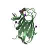
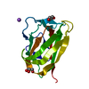
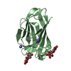
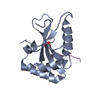
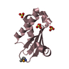
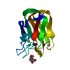
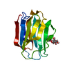
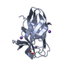
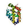
| Title | RNase P protein from Thermotoga maritima in complex with 1-(4-Fluorophenyl)-2-thiourea | ||||||
 Components Components | Ribonuclease P protein component | ||||||
 Keywords Keywords | RNA BINDING PROTEIN | ||||||
| Function / homology |  Function and homology information Function and homology informationtRNA 3'-end processing / 3'-tRNA processing endoribonuclease activity / ribonuclease P complex / ribonuclease P / ribonuclease P activity / tRNA 5'-leader removal / tRNA binding Similarity search - Function | ||||||
| Biological species |   Thermotoga maritima (bacteria) Thermotoga maritima (bacteria) | ||||||
| Method |  X-RAY DIFFRACTION / SYNCHROTRON / MOLECULAR REPLACEMENT / Resolution: 1.54 Å X-RAY DIFFRACTION / SYNCHROTRON / MOLECULAR REPLACEMENT / Resolution: 1.54 Å | ||||||
 Authors Authors | Torres-Larios, A. / Madrigal-Carrillo, E.A. | ||||||
| Funding support |  Mexico, 1items Mexico, 1items
| ||||||
 Citation Citation | Journal: To Be Published Title: Discovery of novel RNase P inhibitors via an activity-binding-structure pipeline Authors: Madrigal-Carrillo, E.A. / Diaz-Tufinio, C.A. / Santamaria-Suarez, H.A. / Arciniega-Castro, M. / Torres-Larios, A. | ||||||
| History |
|
- Structure visualization
| Structure viewer | Molecule: MolmilJmol/JSmol |
|---|
- Downloads & links
-Download
| PDBx/mmCIF format | 6cqc.cif.gz | 290.8 KB | Display | PDBx/mmCIF format |
|---|---|---|---|---|
| PDB format | pdb6cqc.ent.gz | 241.7 KB | Display | PDB format |
| PDBx/mmJSON format | 6cqc.json.gz | Tree view | PDBx/mmJSON format | |
| Others |  Other downloads Other downloads |
-Validation report
| Arichive directory | https://data.pdbj.org/pub/pdb/validation_reports/cq/6cqcftp://data.pdbj.org/pub/pdb/validation_reports/cq/6cqc | HTTPS FTP |
|---|
-Related structure data
| Related structure data | 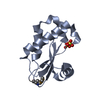 1nz0S S: Starting model for refinement |
|---|---|
| Similar structure data |

-Links
 PDBj
PDBj
- Assembly
| Deposited unit | 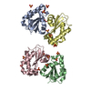
| ||||||||
|---|---|---|---|---|---|---|---|---|---|
| 1 | 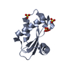
| ||||||||
| 2 | 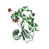
| ||||||||
| 3 | 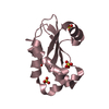
| ||||||||
| 4 | 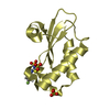
| ||||||||
| Unit cell |
|
-Components
| #1: Protein | Mass: 14363.003 Da / Num. of mol.: 4 Source method: isolated from a genetically manipulated source Source: (gene. exp.) Thermotoga maritima (strain ATCC 43589 / MSB8 / DSM 3109 / JCM 10099) (bacteria)Strain: ATCC 43589 / MSB8 / DSM 3109 / JCM 10099 / Gene: rnpA, TM_1463 / Production host: #2: Chemical | ChemComp-SO4 /   Mass: 96.063 Da / Num. of mol.: 15 / Source method: isolated from a natural source / Formula: SO4 Mass: 96.063 Da / Num. of mol.: 15 / Source method: isolated from a natural source / Formula: SO4#3: Chemical | ChemComp-8P4 / |   Mass: 170.207 Da / Num. of mol.: 1 / Source method: obtained synthetically / Formula: C7H7FN2S Mass: 170.207 Da / Num. of mol.: 1 / Source method: obtained synthetically / Formula: C7H7FN2S#4: Water | ChemComp-HOH / |  Mass: 18.015 Da / Num. of mol.: 486 / Source method: isolated from a natural source / Formula: H2O Mass: 18.015 Da / Num. of mol.: 486 / Source method: isolated from a natural source / Formula: H2O |
|---|
-Experimental details
-Experiment
| Experiment | Method: X-RAY DIFFRACTION / Number of used crystals: 1 |
|---|
- Sample preparation
| Crystal | Density Matthews: 2.09 Å3/Da / Density % sol: 41.21 % |
|---|---|
| Crystal grow | Temperature: 292 K / Method: vapor diffusion, sitting drop / pH: 4.8 Details: P protein from T. maritima in 50 mM Tris-HCl pH 7.5, 0.2 mM EDTA was crystallized at 3 mg/mL in 12% PEG-1000, 100 mm sodium acetate pH 4.8 and 200 mM potassium sulfate |
-Data collection
| Diffraction | Mean temperature: 100 K |
|---|---|
| Diffraction source | Source: SYNCHROTRON / Site: APS  / Beamline: 21-ID-G / Wavelength: 0.9785 Å / Beamline: 21-ID-G / Wavelength: 0.9785 Å |
| Detector | Type: MAR CCD 165 mm / Detector: CCD / Date: Nov 12, 2011 / Details: mirrors |
| Radiation | Monochromator: C(111) / Protocol: SINGLE WAVELENGTH / Monochromatic (M) / Laue (L): M / Scattering type: x-ray |
| Radiation wavelength | Wavelength: 0.9785 Å / Relative weight: 1 |
| Reflection | Resolution: 1.54→46.29 Å / Num. obs: 67977 / % possible obs: 96.9 % / Redundancy: 3.2 % / Biso Wilson estimate: 18 Å2 / CC1/2: 0.993 / Rmerge(I) obs: 0.044 / Rpim(I) all: 0.029 / Rrim(I) all: 0.053 / Χ2: 0.71 / Net I/σ(I): 19.5 |
| Reflection shell | Resolution: 1.54→1.57 Å / Redundancy: 2.9 % / Rmerge(I) obs: 0.237 / Mean I/σ(I) obs: 3.7 / Num. unique obs: 2724 / CC1/2: 0.915 / Rpim(I) all: 0.195 / Rrim(I) all: 0.287 / Χ2: 0.58 / % possible all: 78.5 |
- Processing
| Software |
| ||||||||||||||||||||||||||||||||||||||||||||||||||||||||||||||||||||||||||||||||||||||||||||||||||||||||||||||||||||||||||||||||||||||||||||||||||||||||||||||||||||||||||||||||||||||
|---|---|---|---|---|---|---|---|---|---|---|---|---|---|---|---|---|---|---|---|---|---|---|---|---|---|---|---|---|---|---|---|---|---|---|---|---|---|---|---|---|---|---|---|---|---|---|---|---|---|---|---|---|---|---|---|---|---|---|---|---|---|---|---|---|---|---|---|---|---|---|---|---|---|---|---|---|---|---|---|---|---|---|---|---|---|---|---|---|---|---|---|---|---|---|---|---|---|---|---|---|---|---|---|---|---|---|---|---|---|---|---|---|---|---|---|---|---|---|---|---|---|---|---|---|---|---|---|---|---|---|---|---|---|---|---|---|---|---|---|---|---|---|---|---|---|---|---|---|---|---|---|---|---|---|---|---|---|---|---|---|---|---|---|---|---|---|---|---|---|---|---|---|---|---|---|---|---|---|---|---|---|---|---|
| Refinement | Method to determine structure: MOLECULAR REPLACEMENT Starting model: 1NZ0 Resolution: 1.54→46.286 Å / SU ML: 0.16 / Cross valid method: FREE R-VALUE / σ(F): 1.4 / Phase error: 19.85 / Stereochemistry target values: ML
| ||||||||||||||||||||||||||||||||||||||||||||||||||||||||||||||||||||||||||||||||||||||||||||||||||||||||||||||||||||||||||||||||||||||||||||||||||||||||||||||||||||||||||||||||||||||
| Solvent computation | Shrinkage radii: 0.9 Å / VDW probe radii: 1.11 Å / Solvent model: FLAT BULK SOLVENT MODEL | ||||||||||||||||||||||||||||||||||||||||||||||||||||||||||||||||||||||||||||||||||||||||||||||||||||||||||||||||||||||||||||||||||||||||||||||||||||||||||||||||||||||||||||||||||||||
| Refinement step | Cycle: LAST / Resolution: 1.54→46.286 Å
| ||||||||||||||||||||||||||||||||||||||||||||||||||||||||||||||||||||||||||||||||||||||||||||||||||||||||||||||||||||||||||||||||||||||||||||||||||||||||||||||||||||||||||||||||||||||
| Refine LS restraints |
| ||||||||||||||||||||||||||||||||||||||||||||||||||||||||||||||||||||||||||||||||||||||||||||||||||||||||||||||||||||||||||||||||||||||||||||||||||||||||||||||||||||||||||||||||||||||
| LS refinement shell |
| ||||||||||||||||||||||||||||||||||||||||||||||||||||||||||||||||||||||||||||||||||||||||||||||||||||||||||||||||||||||||||||||||||||||||||||||||||||||||||||||||||||||||||||||||||||||
| Refinement TLS params. | Method: refined / Origin x: 6.7409 Å / Origin y: 13.3899 Å / Origin z: 16.142 Å
| ||||||||||||||||||||||||||||||||||||||||||||||||||||||||||||||||||||||||||||||||||||||||||||||||||||||||||||||||||||||||||||||||||||||||||||||||||||||||||||||||||||||||||||||||||||||
| Refinement TLS group | Selection details: all |
