 Movie
Movie Controller

[English] 日本語
 Yorodumi
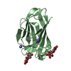
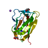
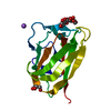
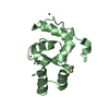
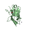
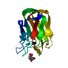
- PDB-1w9s: Structure of a beta-1,3-glucan binding CBM6 from Bacillus halodurans -
+ Open data

- Basic information
| Entry | Database: PDB / ID: 1w9s | ||||||
|---|---|---|---|---|---|---|---|
| Title | Structure of a beta-1,3-glucan binding CBM6 from Bacillus halodurans | ||||||
 Components Components | BH0236 PROTEIN | ||||||
 Keywords Keywords | CARBOHYDRATE-BINDING MODULE / LECTIN / BETA-GLUCAN / CARBOHYDRATE BINDING / GLYCOSIDE HYDROLASE | ||||||
| Function / homology |  Function and homology information Function and homology informationendo-1,3(4)-beta-glucanase activity / glucan endo-1,3-beta-D-glucosidase / glucan endo-1,3-beta-D-glucosidase activity / polysaccharide catabolic process / cell wall organization / carbohydrate binding / extracellular region Similarity search - Function | ||||||
| Biological species |  BACILLUS HALODURANS (bacteria) BACILLUS HALODURANS (bacteria) | ||||||
| Method |  X-RAY DIFFRACTION / MOLECULAR REPLACEMENT / Resolution: 1.59 Å X-RAY DIFFRACTION / MOLECULAR REPLACEMENT / Resolution: 1.59 Å | ||||||
 Authors Authors | Boraston, A.B. / van Bueren, A.L. | ||||||
 Citation Citation | Journal: J.Biol.Chem. / Year: 2005 Title: Family 6 Carbohydrate Binding Modules Recognize the Non-Reducing End of Beta-1,3-Linked Glucans by Presenting a Unique Ligand Binding Surface Authors: Van Bueren, A.L. / Moreland, C. / Gilbert, H.J. / Boraston, A.B. | ||||||
| History |
|
- Structure visualization
| Structure viewer | Molecule: MolmilJmol/JSmol |
|---|
- Downloads & links
-Download
| PDBx/mmCIF format | 1w9s.cif.gz | 133.7 KB | Display | PDBx/mmCIF format |
|---|---|---|---|---|
| PDB format | pdb1w9s.ent.gz | 104 KB | Display | PDB format |
| PDBx/mmJSON format | 1w9s.json.gz | Tree view | PDBx/mmJSON format | |
| Others |  Other downloads Other downloads |
-Validation report
| Arichive directory | https://data.pdbj.org/pub/pdb/validation_reports/w9/1w9sftp://data.pdbj.org/pub/pdb/validation_reports/w9/1w9s | HTTPS FTP |
|---|
-Related structure data
| Related structure data |  1w9tC 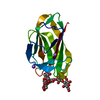 1w9wC 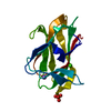 1gmmS C: citing same article ( S: Starting model for refinement |
|---|---|
| Similar structure data |


-Links
 PDBj
PDBj- Assembly
| Deposited unit | 
| ||||||||
|---|---|---|---|---|---|---|---|---|---|
| 1 | 
| ||||||||
| 2 | 
| ||||||||
| Unit cell |
|
-Components
| #1: Protein | Mass: 15673.985 Da / Num. of mol.: 2 / Fragment: CBM, RESIDUES 790-925 Source method: isolated from a genetically manipulated source Source: (gene. exp.) BACILLUS HALODURANS (bacteria) / Production host: #2: Chemical | ChemComp-NA /   Mass: 22.990 Da / Num. of mol.: 6 / Source method: obtained synthetically / Formula: Na Mass: 22.990 Da / Num. of mol.: 6 / Source method: obtained synthetically / Formula: Na#3: Chemical |   Mass: 92.094 Da / Num. of mol.: 2 / Source method: obtained synthetically / Formula: C3H8O3 Mass: 92.094 Da / Num. of mol.: 2 / Source method: obtained synthetically / Formula: C3H8O3#4: Water | ChemComp-HOH / |  Mass: 18.015 Da / Num. of mol.: 350 / Source method: isolated from a natural source / Formula: H2O Mass: 18.015 Da / Num. of mol.: 350 / Source method: isolated from a natural source / Formula: H2O |
|---|
-Experimental details
-Experiment
| Experiment | Method: X-RAY DIFFRACTION |
|---|
- Sample preparation
| Crystal | Density Matthews: 2.04 Å3/Da / Density % sol: 39.63 % |
|---|
-Data collection
| Diffraction | Mean temperature: 113 K |
|---|---|
| Diffraction source | Source: ROTATING ANODE / Wavelength: 1.5418 |
| Detector | Type: RIGAKU RAXIS IV / Detector: IMAGE PLATE |
| Radiation | Protocol: SINGLE WAVELENGTH / Monochromatic (M) / Laue (L): M / Scattering type: x-ray |
| Radiation wavelength | Wavelength: 1.5418 Å / Relative weight: 1 |
| Reflection | Resolution: 1.59→20 Å / Num. obs: 28471 / % possible obs: 90.1 % / Observed criterion σ(I): 2 / Redundancy: 3.7 % / Rmerge(I) obs: 0.03 / Net I/σ(I): 24.9 |
| Reflection shell | Resolution: 1.59→1.65 Å / Redundancy: 3.2 % / Rmerge(I) obs: 0.17 / Mean I/σ(I) obs: 5.7 / % possible all: 68.5 |
- Processing
| Software |
| ||||||||||||||||||||||||||||||||||||||||||||||||||||||||||||||||||||||||||||||||||||||||||||||||||||||||||||||||||||||||||||||||||||||||||||||||||||||||||||||||||||||||||||||||||||||
|---|---|---|---|---|---|---|---|---|---|---|---|---|---|---|---|---|---|---|---|---|---|---|---|---|---|---|---|---|---|---|---|---|---|---|---|---|---|---|---|---|---|---|---|---|---|---|---|---|---|---|---|---|---|---|---|---|---|---|---|---|---|---|---|---|---|---|---|---|---|---|---|---|---|---|---|---|---|---|---|---|---|---|---|---|---|---|---|---|---|---|---|---|---|---|---|---|---|---|---|---|---|---|---|---|---|---|---|---|---|---|---|---|---|---|---|---|---|---|---|---|---|---|---|---|---|---|---|---|---|---|---|---|---|---|---|---|---|---|---|---|---|---|---|---|---|---|---|---|---|---|---|---|---|---|---|---|---|---|---|---|---|---|---|---|---|---|---|---|---|---|---|---|---|---|---|---|---|---|---|---|---|---|---|
| Refinement | Method to determine structure: MOLECULAR REPLACEMENT Starting model: PDB ENTRY 1GMM Resolution: 1.59→20 Å / Cor.coef. Fo:Fc: 0.978 / Cor.coef. Fo:Fc free: 0.96 / SU B: 2.93 / SU ML: 0.048 / Cross valid method: THROUGHOUT / ESU R: 0.127 / ESU R Free: 0.082 / Stereochemistry target values: MAXIMUM LIKELIHOOD / Details: HYDROGENS HAVE BEEN ADDED IN THE RIDING POSITIONS.
| ||||||||||||||||||||||||||||||||||||||||||||||||||||||||||||||||||||||||||||||||||||||||||||||||||||||||||||||||||||||||||||||||||||||||||||||||||||||||||||||||||||||||||||||||||||||
| Solvent computation | Ion probe radii: 0.8 Å / Shrinkage radii: 0.8 Å / VDW probe radii: 1.2 Å / Solvent model: BABINET MODEL WITH MASK | ||||||||||||||||||||||||||||||||||||||||||||||||||||||||||||||||||||||||||||||||||||||||||||||||||||||||||||||||||||||||||||||||||||||||||||||||||||||||||||||||||||||||||||||||||||||
| Displacement parameters | Biso mean: 13.68 Å2
| ||||||||||||||||||||||||||||||||||||||||||||||||||||||||||||||||||||||||||||||||||||||||||||||||||||||||||||||||||||||||||||||||||||||||||||||||||||||||||||||||||||||||||||||||||||||
| Refinement step | Cycle: LAST / Resolution: 1.59→20 Å
| ||||||||||||||||||||||||||||||||||||||||||||||||||||||||||||||||||||||||||||||||||||||||||||||||||||||||||||||||||||||||||||||||||||||||||||||||||||||||||||||||||||||||||||||||||||||
| Refine LS restraints |
|