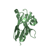
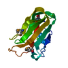
BIOMOLECULE: 1, 2 THIS ENTRY CONTAINS THE CRYSTALLOGRAPHIC ASYMMETRIC UNIT WHICH CONSISTS OF 2 ... BIOMOLECULE: 1, 2 THIS ENTRY CONTAINS THE CRYSTALLOGRAPHIC ASYMMETRIC UNIT WHICH CONSISTS OF 2 CHAIN(S). AUTHORS STATE THAT THE BIOLOGICAL UNIT OF THIS PROTEIN IS UNKNOWN.
Resolution: 1.9→20 Å / Cor.coef. Fo:Fc: 0.952 / Cor.coef. Fo:Fc free: 0.928 / WRfactor Rfree: 0.238 / WRfactor Rwork: 0.185 / SU B: 3.872 / SU ML: 0.117 / Cross valid method: FREE R / ESU R: 0.195 / ESU R Free: 0.174 / Stereochemistry target values: MAXIMUM LIKELIHOOD Details: HYDROGENS HAVE BEEN ADDED IN THE RIDING POSITIONS. Arp/warp, molprobity, coot have also been used in structure solution and refinement
Rfactor
Num. reflection
% reflection
Selection details
Rfree
0.2457
998
5.995 %
thin shells
Rwork
0.1915
-
-
-
obs
0.195
16648
94.334 %
-
Solvent computation
Ion probe radii: 0.8 Å / Shrinkage radii: 0.8 Å / VDW probe radii: 1.4 Å / Solvent model: MASK BULK SOLVENT
Displacement parameters
Biso mean: 20.834 Å2
Baniso -1
Baniso -2
Baniso -3
1-
0.336 Å2
-0.612 Å2
0.204 Å2
2-
-
0.198 Å2
-0.296 Å2
3-
-
-
-0.722 Å2
Refinement step
Cycle: LAST / Resolution: 1.9→20 Å
Protein
Nucleic acid
Ligand
Solvent
Total
Num. atoms
1941
0
11
65
2017
Refine LS restraints
Refine-ID
Type
Dev ideal
Dev ideal target
Number
X-RAY DIFFRACTION
r_bond_refined_d
0.017
0.022
1998
X-RAY DIFFRACTION
r_bond_other_d
0.002
0.02
1386
X-RAY DIFFRACTION
r_angle_refined_deg
1.358
1.942
2693
X-RAY DIFFRACTION
r_angle_other_deg
0.956
3.006
3347
X-RAY DIFFRACTION
r_dihedral_angle_1_deg
6.342
5
245
X-RAY DIFFRACTION
r_dihedral_angle_2_deg
32.238
24.314
102
X-RAY DIFFRACTION
r_dihedral_angle_3_deg
12.665
15
349
X-RAY DIFFRACTION
r_dihedral_angle_4_deg
16.666
15
15
X-RAY DIFFRACTION
r_chiral_restr
0.085
0.2
281
X-RAY DIFFRACTION
r_gen_planes_refined
0.005
0.02
2237
X-RAY DIFFRACTION
r_gen_planes_other
0.001
0.02
412
X-RAY DIFFRACTION
r_nbd_refined
0.198
0.2
381
X-RAY DIFFRACTION
r_nbd_other
0.189
0.2
1482
X-RAY DIFFRACTION
r_nbtor_refined
0.172
0.2
958
X-RAY DIFFRACTION
r_nbtor_other
0.084
0.2
979
X-RAY DIFFRACTION
r_xyhbond_nbd_refined
0.137
0.2
75
X-RAY DIFFRACTION
r_symmetry_vdw_refined
0.131
0.2
7
X-RAY DIFFRACTION
r_symmetry_vdw_other
0.208
0.2
39
X-RAY DIFFRACTION
r_symmetry_hbond_refined
0.098
0.2
5
X-RAY DIFFRACTION
r_mcbond_it
2.717
2
1359
X-RAY DIFFRACTION
r_mcbond_other
0.797
2
491
X-RAY DIFFRACTION
r_mcangle_it
3.206
3
1933
X-RAY DIFFRACTION
r_scbond_it
2.471
2
885
X-RAY DIFFRACTION
r_scangle_it
3.527
3
757
LS refinement shell
Refine-ID: X-RAY DIFFRACTION / Total num. of bins used: 20
Resolution (Å)
Rfactor Rfree
Num. reflection Rfree
Rfactor Rwork
Num. reflection Rwork
Rfactor all
Num. reflection all
% reflection obs (%)
1.9-1.949
0.367
62
0.249
1142
0.256
1331
90.458
1.949-2.002
0.322
78
0.217
1062
0.224
1241
91.861
2.002-2.06
0.336
102
0.22
1032
0.23
1234
91.896
2.06-2.123
0
0.207
1100
0.207
1197
91.896
2.123-2.192
0.272
102
0.197
983
0.205
1169
92.814
2.192-2.269
0
0.209
1053
0.209
1121
93.934
2.269-2.354
0.274
107
0.204
916
0.211
1094
93.51
2.354-2.45
0.258
92
0.192
887
0.198
1043
93.864
2.45-2.558
0
0.187
936
0.187
987
94.833
2.558-2.682
0.251
81
0.203
845
0.207
972
95.267
2.682-2.825
0.262
76
0.197
788
0.203
905
95.47
2.825-2.995
0
0.216
820
0.216
855
95.906
2.995-3.2
0.258
62
0.202
742
0.206
832
96.635
3.2-3.453
0.26
50
0.186
665
0.191
738
96.883
3.453-3.777
0.212
43
0.172
633
0.174
693
97.547
3.777-4.215
0.186
40
0.158
563
0.16
618
97.573
4.215-4.851
0.175
28
0.145
530
0.147
568
98.239
4.851-5.903
0.224
40
0.192
425
0.195
472
98.517
5.903-8.191
0.235
20
0.21
337
0.212
360
99.167
8.191-30
0.222
15
0.201
191
0.203
218
94.495
+
About Yorodumi
-
News
-
Feb 9, 2022. New format data for meta-information of EMDB entries
New format data for meta-information of EMDB entries
Version 3 of the EMDB header file is now the official format.
The previous official version 1.9 will be removed from the archive.
In the structure databanks used in Yorodumi, some data are registered as the other names, "COVID-19 virus" and "2019-nCoV". Here are the details of the virus and the list of structure data.
Jan 31, 2019. EMDB accession codes are about to change! (news from PDBe EMDB page)
EMDB accession codes are about to change! (news from PDBe EMDB page)
The allocation of 4 digits for EMDB accession codes will soon come to an end. Whilst these codes will remain in use, new EMDB accession codes will include an additional digit and will expand incrementally as the available range of codes is exhausted. The current 4-digit format prefixed with “EMD-” (i.e. EMD-XXXX) will advance to a 5-digit format (i.e. EMD-XXXXX), and so on. It is currently estimated that the 4-digit codes will be depleted around Spring 2019, at which point the 5-digit format will come into force.
The EM Navigator/Yorodumi systems omit the EMD- prefix.
Related info.:Q: What is EMD? / ID/Accession-code notation in Yorodumi/EM Navigator
Yorodumi is a browser for structure data from EMDB, PDB, SASBDB, etc.
This page is also the successor to EM Navigator detail page, and also detail information page/front-end page for Omokage search.
The word "yorodu" (or yorozu) is an old Japanese word meaning "ten thousand". "mi" (miru) is to see.
Related info.:EMDB / PDB / SASBDB / Comparison of 3 databanks / Yorodumi Search / Aug 31, 2016. New EM Navigator & Yorodumi / Yorodumi Papers / Jmol/JSmol / Function and homology information / Changes in new EM Navigator and Yorodumi
 Movie
Movie Controller

 Yorodumi
 Open data
 Basic information
 Components
 Keywords
 Function and homology information
 Homo sapiens (human)
 X-RAY DIFFRACTION /
  Authors
 Citation
 Structure visualization
 Downloads & links
 Other downloads
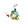


 PDBj
PDBj


 Assembly
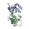


 Mass: 65.409 Da / Num. of mol.: 2 / Source method: obtained synthetically / Formula: Zn
 Mass: 78.133 Da / Num. of mol.: 2 / Source method: obtained synthetically / Formula: C2H6OS
 Num. of mol.: 3 / Source method: obtained synthetically
 Mass: 18.015 Da / Num. of mol.: 65 / Source method: isolated from a natural source / Formula: H2O
 Sample preparation
 Processing