 Movie
Movie Controller


+ Open data

- Basic information
| Entry | Database: PDB / ID: 6cq3 | ||||||||||||||||||||||||||||
|---|---|---|---|---|---|---|---|---|---|---|---|---|---|---|---|---|---|---|---|---|---|---|---|---|---|---|---|---|---|
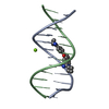
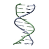
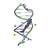
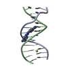
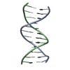
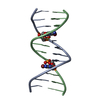
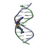
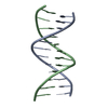
| Title | Crystal structure of DNA dodecamer D(CGCGAATTCGCG) | ||||||||||||||||||||||||||||
 Components Components | DNA (5'-D(* Keywords KeywordsDNA / dodecamer | Function / homology | : / DNA / DNA (> 10) |  Function and homology information Function and homology informationBiological species | synthetic construct (others) | Method |  X-RAY DIFFRACTION / SYNCHROTRON / MOLECULAR REPLACEMENT / Resolution: 1.3 Å X-RAY DIFFRACTION / SYNCHROTRON / MOLECULAR REPLACEMENT / Resolution: 1.3 Å  Authors AuthorsDelgado, J.L. / Vance, N.R. / Kerns, R.J. | Funding support | |  United States, 1items United States, 1items
 CitationJournal: To Be Published CitationJournal: To Be PublishedTitle: Crystal structure of DNA dodecamer D(CGCGAATTCGCG) Authors: Delgado, J.L. / Vance, N.R. / Kerns, R.J. History |
|
- Structure visualization
| Structure viewer | Molecule: MolmilJmol/JSmol |
|---|
- Downloads & links
-Download
| PDBx/mmCIF format | 6cq3.cif.gz | 51.1 KB | Display | PDBx/mmCIF format |
|---|---|---|---|---|
| PDB format | pdb6cq3.ent.gz | 37 KB | Display | PDB format |
| PDBx/mmJSON format | 6cq3.json.gz | Tree view | PDBx/mmJSON format | |
| Others |  Other downloads Other downloads |
-Validation report
| Arichive directory | https://data.pdbj.org/pub/pdb/validation_reports/cq/6cq3ftp://data.pdbj.org/pub/pdb/validation_reports/cq/6cq3 | HTTPS FTP |
|---|
-Related structure data
| Related structure data | 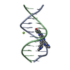 8bnaS S: Starting model for refinement |
|---|---|
| Similar structure data |


-Links
 PDBj
PDBj


- Assembly
| Deposited unit | 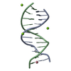
| ||||||||
|---|---|---|---|---|---|---|---|---|---|
| 1 |
| ||||||||
| Unit cell |
|
-Components
| #1: DNA chain | Mass: 3663.392 Da / Num. of mol.: 2 / Source method: obtained synthetically / Source: (synth.) synthetic construct (others) #2: Chemical |   Mass: 24.305 Da / Num. of mol.: 3 / Source method: obtained synthetically / Formula: Mg Mass: 24.305 Da / Num. of mol.: 3 / Source method: obtained synthetically / Formula: Mg#3: Chemical | ChemComp-CO / |   Mass: 58.933 Da / Num. of mol.: 1 / Source method: obtained synthetically / Formula: Co Mass: 58.933 Da / Num. of mol.: 1 / Source method: obtained synthetically / Formula: Co#4: Water | ChemComp-HOH / |  Mass: 18.015 Da / Num. of mol.: 173 / Source method: isolated from a natural source / Formula: H2O Mass: 18.015 Da / Num. of mol.: 173 / Source method: isolated from a natural source / Formula: H2O |
|---|
-Experimental details
-Experiment
| Experiment | Method: X-RAY DIFFRACTION / Number of used crystals: 1 |
|---|
- Sample preparation
| Crystal | Density Matthews: 2.19 Å3/Da / Density % sol: 43.76 % |
|---|---|
| Crystal grow | Temperature: 277 K / Method: vapor diffusion, hanging drop / pH: 5.5 Details: 10% (v/v) 2-Methyl-2,4-pentanediol, 40mM Sodium cacodylate trihydrate, 20mM Hexammine cobalt(III) chloride, 40mM Lithium chloride, 20mM Magnesium chloride hexahydrate |
-Data collection
| Diffraction | Mean temperature: 100 K |
|---|---|
| Diffraction source | Source: SYNCHROTRON / Site: ALS / Beamline: 4.2.2 / Wavelength: 1.0008 Å |
| Detector | Type: RDI CMOS_8M / Detector: CMOS / Date: Jan 25, 2018 |
| Radiation | Monochromator: Si(111) / Protocol: SINGLE WAVELENGTH / Monochromatic (M) / Laue (L): M / Scattering type: x-ray |
| Radiation wavelength | Wavelength: 1.0008 Å / Relative weight: 1 |
| Reflection | Resolution: 1.3→34.384 Å / Num. obs: 16300 / % possible obs: 99.04 % / Redundancy: 9.6 % / CC1/2: 1 / Rmerge(I) obs: 0.047 / Rpim(I) all: 0.021 / Rrim(I) all: 0.054 / Rsym value: 0.052 / Net I/σ(I): 25.8 |
| Reflection shell | Resolution: 1.3→1.32 Å / Redundancy: 7.1 % / Rmerge(I) obs: 0.753 / Mean I/σ(I) obs: 2.4 / Num. unique obs: 47 / CC1/2: 0.779 / Rpim(I) all: 0.446 / Rrim(I) all: 0.887 / Rsym value: 0.815 / % possible all: 98.7 |
- Processing
| Software |
| |||||||||||||||||||||||||||||||||||||||||||||||||
|---|---|---|---|---|---|---|---|---|---|---|---|---|---|---|---|---|---|---|---|---|---|---|---|---|---|---|---|---|---|---|---|---|---|---|---|---|---|---|---|---|---|---|---|---|---|---|---|---|---|---|
| Refinement | Method to determine structure: MOLECULAR REPLACEMENT Starting model: 8BNA Resolution: 1.3→34.384 Å / SU ML: 0.12 / Cross valid method: FREE R-VALUE / σ(F): 1.34 / Phase error: 19.13 / Stereochemistry target values: ML
| |||||||||||||||||||||||||||||||||||||||||||||||||
| Solvent computation | Shrinkage radii: 0.9 Å / VDW probe radii: 1.11 Å / Solvent model: FLAT BULK SOLVENT MODEL | |||||||||||||||||||||||||||||||||||||||||||||||||
| Refinement step | Cycle: LAST / Resolution: 1.3→34.384 Å
| |||||||||||||||||||||||||||||||||||||||||||||||||
| Refine LS restraints |
| |||||||||||||||||||||||||||||||||||||||||||||||||
| LS refinement shell |
|
