 Movie
Movie Controller

[English] 日本語
 Yorodumi
- PDB-6ci5: Crystal structure of the formyltransferase PseJ from Anoxybacillu... -
+ Open data

- Basic information
| Entry | Database: PDB / ID: 6ci5 | ||||||
|---|---|---|---|---|---|---|---|
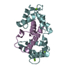
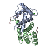
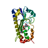
| Title | Crystal structure of the formyltransferase PseJ from Anoxybacillus kamchatkensis in complex with UDP-4,6-dideoxy-4-formamido-L-AltNAc and tetrahydrofolate | ||||||
 Components Components | formyltransferase PseJ | ||||||
 Keywords Keywords | TRANSFERASE / formyltransferase | ||||||
| Function / homology | Chem-1YJ / Chem-F5G Function and homology information Function and homology information | ||||||
| Biological species |  Anoxybacillus kamchatkensis G10 (bacteria) Anoxybacillus kamchatkensis G10 (bacteria) | ||||||
| Method |  X-RAY DIFFRACTION / SYNCHROTRON / MOLECULAR REPLACEMENT / Resolution: 2.00003052726 Å X-RAY DIFFRACTION / SYNCHROTRON / MOLECULAR REPLACEMENT / Resolution: 2.00003052726 Å | ||||||
 Authors Authors | Reimer, J.M. / Harb, I. / Schmeing, T.M. | ||||||
| Funding support |  Canada, 1items Canada, 1items
| ||||||
 Citation Citation | Journal: ACS Chem. Biol. / Year: 2018 Title: Structural Insight into a Novel Formyltransferase and Evolution to a Nonribosomal Peptide Synthetase Tailoring Domain. Authors: Reimer, J.M. / Harb, I. / Ovchinnikova, O.G. / Jiang, J. / Whitfield, C. / Schmeing, T.M. | ||||||
| History |
|
- Structure visualization
| Structure viewer | Molecule: MolmilJmol/JSmol |
|---|
- Downloads & links
-Download
| PDBx/mmCIF format | 6ci5.cif.gz | 156.6 KB | Display | PDBx/mmCIF format |
|---|---|---|---|---|
| PDB format | pdb6ci5.ent.gz | 105 KB | Display | PDB format |
| PDBx/mmJSON format | 6ci5.json.gz | Tree view | PDBx/mmJSON format | |
| Others |  Other downloads Other downloads |
-Validation report
| Summary document | 6ci5_validation.pdf.gz | 1.1 MB | Display | wwPDB validaton report |
|---|---|---|---|---|
| Full document | 6ci5_full_validation.pdf.gz | 1.1 MB | Display | |
| Data in XML | 6ci5_validation.xml.gz | 10.4 KB | Display | |
| Data in CIF | 6ci5_validation.cif.gz | 13.1 KB | Display | |
| Arichive directory | https://data.pdbj.org/pub/pdb/validation_reports/ci/6ci5ftp://data.pdbj.org/pub/pdb/validation_reports/ci/6ci5 | HTTPS FTP |
-Related structure data
| Related structure data | 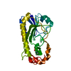 6ci2SC  6ci4C 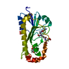 6edkC S: Starting model for refinement C: citing same article ( |
|---|---|
| Similar structure data |


-Links
 PDBj
PDBj- Assembly
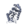
| Deposited unit | 
| ||||||||||||
|---|---|---|---|---|---|---|---|---|---|---|---|---|---|
| 1 |
| ||||||||||||
| Unit cell |
|
-Components
| #1: Protein | Mass: 25609.049 Da / Num. of mol.: 1 Source method: isolated from a genetically manipulated source Source: (gene. exp.) Anoxybacillus kamchatkensis G10 (bacteria)Production host: |
|---|---|
| #2: Chemical | ChemComp-SO4 /   Mass: 96.063 Da / Num. of mol.: 1 / Source method: isolated from a natural source / Formula: SO4 Mass: 96.063 Da / Num. of mol.: 1 / Source method: isolated from a natural source / Formula: SO4 |
| #3: Chemical | ChemComp-F5G / (  Mass: 618.380 Da / Num. of mol.: 1 / Source method: obtained synthetically / Formula: C18H28N4O16P2 Mass: 618.380 Da / Num. of mol.: 1 / Source method: obtained synthetically / Formula: C18H28N4O16P2 |
| #4: Chemical | ChemComp-1YJ /   Mass: 445.429 Da / Num. of mol.: 1 / Source method: obtained synthetically / Formula: C19H23N7O6 Mass: 445.429 Da / Num. of mol.: 1 / Source method: obtained synthetically / Formula: C19H23N7O6 |
| #5: Water | ChemComp-HOH /  Mass: 18.015 Da / Num. of mol.: 31 / Source method: isolated from a natural source / Formula: H2O Mass: 18.015 Da / Num. of mol.: 31 / Source method: isolated from a natural source / Formula: H2O |
-Experimental details
-Experiment
| Experiment | Method: X-RAY DIFFRACTION / Number of used crystals: 1 |
|---|
- Sample preparation
| Crystal | Density Matthews: 2.85 Å3/Da / Density % sol: 56.84 % |
|---|---|
| Crystal grow | Temperature: 295 K / Method: vapor diffusion, sitting drop Details: 0.2 M ammonium sulfate, 0.1 M MES pH 6.5, 25.6% PEG5000 MME |
-Data collection
| Diffraction | Mean temperature: 100 K |
|---|---|
| Diffraction source | Source: SYNCHROTRON / Site: CLSI / Beamline: 08ID-1 / Wavelength: 0.979 Å |
| Detector | Type: DECTRIS PILATUS3 S 6M / Detector: PIXEL / Date: Oct 26, 2017 |
| Radiation | Protocol: SINGLE WAVELENGTH / Monochromatic (M) / Laue (L): M / Scattering type: x-ray |
| Radiation wavelength | Wavelength: 0.979 Å / Relative weight: 1 |
| Reflection | Resolution: 1.7→47.23 Å / Num. obs: 32206 / % possible obs: 100 % / Redundancy: 6.4 % / Biso Wilson estimate: 48.8393855317 Å2 / Rmerge(I) obs: 0.042 / Net I/σ(I): 11.4 |
| Reflection shell | Resolution: 1.9→1.94 Å / Rmerge(I) obs: 0.984 / Num. unique obs: 1720 |
- Processing
| Software |
| |||||||||||||||||||||||||||||||||||||||||||||||||||||||||||||||||||||||||||||||||||||||||||||||||||||||||
|---|---|---|---|---|---|---|---|---|---|---|---|---|---|---|---|---|---|---|---|---|---|---|---|---|---|---|---|---|---|---|---|---|---|---|---|---|---|---|---|---|---|---|---|---|---|---|---|---|---|---|---|---|---|---|---|---|---|---|---|---|---|---|---|---|---|---|---|---|---|---|---|---|---|---|---|---|---|---|---|---|---|---|---|---|---|---|---|---|---|---|---|---|---|---|---|---|---|---|---|---|---|---|---|---|---|---|
| Refinement | Method to determine structure: MOLECULAR REPLACEMENT Starting model: 6CI2 Resolution: 2.00003052726→46.8838402268 Å / SU ML: 0.312089467255 / Cross valid method: FREE R-VALUE / σ(F): 1.47364596753 / Phase error: 30.9228387233 Stereochemistry target values: GeoStd + Monomer Library + CDL v1.2
| |||||||||||||||||||||||||||||||||||||||||||||||||||||||||||||||||||||||||||||||||||||||||||||||||||||||||
| Solvent computation | Shrinkage radii: 0.9 Å / VDW probe radii: 1.11 Å / Solvent model: FLAT BULK SOLVENT MODEL | |||||||||||||||||||||||||||||||||||||||||||||||||||||||||||||||||||||||||||||||||||||||||||||||||||||||||
| Displacement parameters | Biso mean: 74.0064049426 Å2 | |||||||||||||||||||||||||||||||||||||||||||||||||||||||||||||||||||||||||||||||||||||||||||||||||||||||||
| Refinement step | Cycle: LAST / Resolution: 2.00003052726→46.8838402268 Å
| |||||||||||||||||||||||||||||||||||||||||||||||||||||||||||||||||||||||||||||||||||||||||||||||||||||||||
| Refine LS restraints |
| |||||||||||||||||||||||||||||||||||||||||||||||||||||||||||||||||||||||||||||||||||||||||||||||||||||||||
| LS refinement shell |
| |||||||||||||||||||||||||||||||||||||||||||||||||||||||||||||||||||||||||||||||||||||||||||||||||||||||||
| Refinement TLS params. | Method: refined / Refine-ID: X-RAY DIFFRACTION
| |||||||||||||||||||||||||||||||||||||||||||||||||||||||||||||||||||||||||||||||||||||||||||||||||||||||||
| Refinement TLS group |
|
