| Entry | Database: PDB / ID: 6cgh
|
|---|
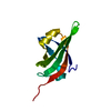
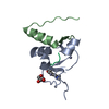
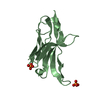
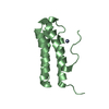
| Title | Solution structure of the four-helix bundle region of human J-protein Zuotin, a component of ribosome-associated complex (RAC) |
|---|
 Components Components | DnaJ homolog subfamily C member 2 |
|---|
 Keywords Keywords | CHAPERONE / Zuotin / J-protein / Hsp70 / molecular chaperone / DNAJC2 / Hsp40 / Ribosome-Associated Complex (RAC) / Mpp11 |
|---|
| Function / homology |  Function and homology information Function and homology information
'de novo' cotranslational protein folding / ubiquitin-modified histone reader activity / ATPase activator activity / Regulation of HSF1-mediated heat shock response / regulation of cellular response to heat / regulation of translational fidelity / Hsp70 protein binding / ribosome binding / protein folding / histone binding ...'de novo' cotranslational protein folding / ubiquitin-modified histone reader activity / ATPase activator activity / Regulation of HSF1-mediated heat shock response / regulation of cellular response to heat / regulation of translational fidelity / Hsp70 protein binding / ribosome binding / protein folding / histone binding / chromatin binding / nucleolus / positive regulation of DNA-templated transcription / DNA-templated transcription / RNA binding / nucleoplasm / nucleus / cytosol / cytoplasmSimilarity search - Function Myb-like DNA-binding domain / Ribosome-associated complex head domain / Ribosome-associated complex head domain superfamily / J-protein Zuotin/DnaJC2 / : / Ribosome-associated complex head domain / Zuotin-like, zuotin homology domain / Nt-dnaJ domain signature. / DnaJ domain, conserved site / DnaJ domain ...Myb-like DNA-binding domain / Ribosome-associated complex head domain / Ribosome-associated complex head domain superfamily / J-protein Zuotin/DnaJC2 / : / Ribosome-associated complex head domain / Zuotin-like, zuotin homology domain / Nt-dnaJ domain signature. / DnaJ domain, conserved site / DnaJ domain / DnaJ molecular chaperone homology domain / dnaJ domain profile. / Chaperone J-domain superfamily / DnaJ domain / SANT domain profile. / SANT domain / Myb domain / SANT SWI3, ADA2, N-CoR and TFIIIB'' DNA-binding domains / SANT/Myb domain / Homeobox-like domain superfamilySimilarity search - Domain/homology |
|---|
| Biological species |  Homo sapiens (human) Homo sapiens (human) |
|---|
| Method | SOLUTION NMR / molecular dynamics |
|---|
 Authors Authors | Shrestha, O.K. / Lee, W. / Tonelli, M. / Cornilescu, G. / Markley, J.L. / Ciesielski, S.J. / Craig, E.A. |
|---|
| Funding support |  United States, 3items United States, 3items | Organization | Grant number | Country |
|---|
| National Institutes of Health/National Human Genome Research Institute (NIH/NHGRI) | GM31107 | United States | | National Institutes of Health/National Human Genome Research Institute (NIH/NHGRI) | GM27870 | United States | | National Institutes of Health/National Human Genome Research Institute (NIH/NHGRI) | P41GM103399 | United States |
|
|---|
 Citation Citation | Journal: Plos One / Year: 2019
Title: Structure and evolution of the 4-helix bundle domain of Zuotin, a J-domain protein co-chaperone of Hsp70.
Authors: Shrestha, O.K. / Sharma, R. / Tomiczek, B. / Lee, W. / Tonelli, M. / Cornilescu, G. / Stolarska, M. / Nierzwicki, L. / Czub, J. / Markley, J.L. / Marszalek, J. / Ciesielski, S.J. / Craig, E.A. |
|---|
| History | | Deposition | Feb 20, 2018 | Deposition site: RCSB / Processing site: RCSB |
|---|
| Revision 1.0 | Jun 12, 2019 | Provider: repository / Type: Initial release |
|---|
| Revision 1.1 | Dec 18, 2019 | Group: Author supporting evidence / Data collection / Category: pdbx_audit_support / pdbx_nmr_spectrometer
Item: _pdbx_audit_support.funding_organization / _pdbx_nmr_spectrometer.model |
|---|
| Revision 1.2 | Jun 14, 2023 | Group: Database references / Other / Category: database_2 / pdbx_database_status
Item: _database_2.pdbx_DOI / _database_2.pdbx_database_accession / _pdbx_database_status.status_code_nmr_data |
|---|
| Revision 1.3 | May 15, 2024 | Group: Data collection / Database references / Category: chem_comp_atom / chem_comp_bond / database_2 / Item: _database_2.pdbx_DOI |
|---|
|
|---|
 Movie
Movie Controller

 Yorodumi
 Open data
 Basic information
 Structure visualization
 Downloads & links
 Other downloads
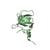


 PDBj
PDBj


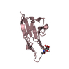
 Assembly
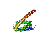
 gel filtration
 Sample preparation