 Movie
Movie Controller


+ Open data

- Basic information
| Entry | Database: PDB / ID: 6c6l | |||||||||||||||||||||||||||||||||||||||||||||||||||||||||||||||||||||||||||||||||||||||||||||||||||||||||||||||||||||
|---|---|---|---|---|---|---|---|---|---|---|---|---|---|---|---|---|---|---|---|---|---|---|---|---|---|---|---|---|---|---|---|---|---|---|---|---|---|---|---|---|---|---|---|---|---|---|---|---|---|---|---|---|---|---|---|---|---|---|---|---|---|---|---|---|---|---|---|---|---|---|---|---|---|---|---|---|---|---|---|---|---|---|---|---|---|---|---|---|---|---|---|---|---|---|---|---|---|---|---|---|---|---|---|---|---|---|---|---|---|---|---|---|---|---|---|---|---|---|
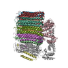
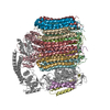
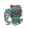
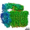
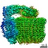
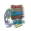
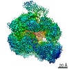
| Title | Yeast Vacuolar ATPase Vo in lipid nanodisc | |||||||||||||||||||||||||||||||||||||||||||||||||||||||||||||||||||||||||||||||||||||||||||||||||||||||||||||||||||||
 Components Components |
| |||||||||||||||||||||||||||||||||||||||||||||||||||||||||||||||||||||||||||||||||||||||||||||||||||||||||||||||||||||
 Keywords Keywords | MEMBRANE PROTEIN / Vacuolar H+-ATPase / Vo proton channel / rotary motor enzyme | |||||||||||||||||||||||||||||||||||||||||||||||||||||||||||||||||||||||||||||||||||||||||||||||||||||||||||||||||||||
| Function / homology |  Function and homology information Function and homology informationcell wall mannoprotein biosynthetic process / ATPase-coupled ion transmembrane transporter activity / protein localization to vacuolar membrane / cellular response to alkaline pH / Insulin receptor recycling / Transferrin endocytosis and recycling / polyphosphate metabolic process / ROS and RNS production in phagocytes / Golgi lumen acidification / Amino acids regulate mTORC1 ...cell wall mannoprotein biosynthetic process / ATPase-coupled ion transmembrane transporter activity / protein localization to vacuolar membrane / cellular response to alkaline pH / Insulin receptor recycling / Transferrin endocytosis and recycling / polyphosphate metabolic process / ROS and RNS production in phagocytes / Golgi lumen acidification / Amino acids regulate mTORC1 / P-type proton-exporting transporter activity / vacuolar transport / vacuolar proton-transporting V-type ATPase, V0 domain / endosomal lumen acidification / vacuole organization / proton-transporting V-type ATPase complex / protein targeting to vacuole / fungal-type vacuole / vacuolar proton-transporting V-type ATPase complex / cellular hyperosmotic response / vacuolar acidification / fungal-type vacuole membrane / phosphatidylinositol-3,5-bisphosphate binding / proton transmembrane transporter activity / proton-transporting ATPase activity, rotational mechanism / intracellular copper ion homeostasis / Neutrophil degranulation / RNA endonuclease activity / proton transmembrane transport / cell periphery / transmembrane transport / endocytosis / ATPase binding / protein-containing complex assembly / intracellular iron ion homeostasis / membrane raft / Golgi membrane / endoplasmic reticulum membrane / membrane Similarity search - Function | |||||||||||||||||||||||||||||||||||||||||||||||||||||||||||||||||||||||||||||||||||||||||||||||||||||||||||||||||||||
| Biological species |  | |||||||||||||||||||||||||||||||||||||||||||||||||||||||||||||||||||||||||||||||||||||||||||||||||||||||||||||||||||||
| Method | ELECTRON MICROSCOPY / single particle reconstruction / cryo EM / Resolution: 3.5 Å | |||||||||||||||||||||||||||||||||||||||||||||||||||||||||||||||||||||||||||||||||||||||||||||||||||||||||||||||||||||
 Authors Authors | Roh, S. / Stam, N.J. / Hryc, C. / Couoh-Cardel, S. / Pintilie, G. / Chiu, W. / Wilkens, S. | |||||||||||||||||||||||||||||||||||||||||||||||||||||||||||||||||||||||||||||||||||||||||||||||||||||||||||||||||||||
| Funding support |  United States, 4items United States, 4items
| |||||||||||||||||||||||||||||||||||||||||||||||||||||||||||||||||||||||||||||||||||||||||||||||||||||||||||||||||||||
 Citation Citation | Journal: Mol Cell / Year: 2018 Title: The 3.5-Å CryoEM Structure of Nanodisc-Reconstituted Yeast Vacuolar ATPase V Proton Channel. Authors: Soung-Hun Roh / Nicholas J Stam / Corey F Hryc / Sergio Couoh-Cardel / Grigore Pintilie / Wah Chiu / Stephan Wilkens / Abstract: The molecular mechanism of transmembrane proton translocation in rotary motor ATPases is not fully understood. Here, we report the 3.5-Å resolution cryoEM structure of the lipid nanodisc- ...The molecular mechanism of transmembrane proton translocation in rotary motor ATPases is not fully understood. Here, we report the 3.5-Å resolution cryoEM structure of the lipid nanodisc-reconstituted V proton channel of the yeast vacuolar H-ATPase, captured in a physiologically relevant, autoinhibited state. The resulting atomic model provides structural detail for the amino acids that constitute the proton pathway at the interface of the proteolipid ring and subunit a. Based on the structure and previous mutagenesis studies, we propose the chemical basis of transmembrane proton transport. Moreover, we discovered that the C terminus of the assembly factor Voa1 is an integral component of mature V. Voa1's C-terminal transmembrane α helix is bound inside the proteolipid ring, where it contributes to the stability of the complex. Our structure rationalizes possible mechanisms by which mutations in human V can result in disease phenotypes and may thus provide new avenues for therapeutic interventions. | |||||||||||||||||||||||||||||||||||||||||||||||||||||||||||||||||||||||||||||||||||||||||||||||||||||||||||||||||||||
| History |
|
- Structure visualization
| Movie |
Movie viewer |
|---|---|
| Structure viewer | Molecule: MolmilJmol/JSmol |
- Downloads & links
-Download
| PDBx/mmCIF format | 6c6l.cif.gz | 556.2 KB | Display | PDBx/mmCIF format |
|---|---|---|---|---|
| PDB format | pdb6c6l.ent.gz | 450.9 KB | Display | PDB format |
| PDBx/mmJSON format | 6c6l.json.gz | Tree view | PDBx/mmJSON format | |
| Others |  Other downloads Other downloads |
-Validation report
| Arichive directory | https://data.pdbj.org/pub/pdb/validation_reports/c6/6c6lftp://data.pdbj.org/pub/pdb/validation_reports/c6/6c6l | HTTPS FTP |
|---|
-Related structure data
| Related structure data |  7348MC M: map data used to model this data C: citing same article ( |
|---|---|
| Similar structure data |

-Links
 PDBj
PDBj


- Assembly
| Deposited unit | 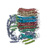
|
|---|---|
| 1 |
|
-Components
-V-type proton ATPase subunit ... , 7 types, 14 molecules DCMEFGHIJKLBAO
| #1: Protein | Mass: 17046.361 Da / Num. of mol.: 1 / Source method: isolated from a natural source Source: (natural) Strain: ATCC 204508 / S288c / References: UniProt: P32842 | ||||||
|---|---|---|---|---|---|---|---|
| #2: Protein | Mass: 22610.641 Da / Num. of mol.: 1 / Source method: isolated from a natural source Source: (natural) Strain: ATCC 204508 / S288c / References: UniProt: P23968 | ||||||
| #4: Protein | Mass: 8387.065 Da / Num. of mol.: 1 / Source method: isolated from a natural source Source: (natural) Strain: ATCC 204508 / S288c / References: UniProt: Q3E7B6 | ||||||
| #5: Protein | Mass: 16357.501 Da / Num. of mol.: 8 / Source method: isolated from a natural source Source: (natural) Strain: ATCC 204508 / S288c References: UniProt: P25515, H+-transporting two-sector ATPase #6: Protein | | Mass: 39822.484 Da / Num. of mol.: 1 / Source method: isolated from a natural source Source: (natural) Strain: ATCC 204508 / S288c / References: UniProt: P32366 #7: Protein | | Mass: 95625.484 Da / Num. of mol.: 1 / Mutation: C-terminal calmodulin binding peptide / Source method: isolated from a natural source Source: (natural) Strain: ATCC 204508 / S288c / References: UniProt: P32563 #8: Protein | | Mass: 9369.934 Da / Num. of mol.: 1 / Source method: isolated from a natural source Source: (natural) Strain: ATCC 204508 / S288c / References: UniProt: P0C5R9 |
-Protein , 1 types, 1 molecules N
| #3: Protein | Mass: 29694.885 Da / Num. of mol.: 1 / Source method: isolated from a natural source Source: (natural) Strain: ATCC 204508 / S288c / References: UniProt: P53262 |
|---|
-Details
| Has protein modification | N |
|---|
-Experimental details
-Experiment
| Experiment | Method: ELECTRON MICROSCOPY |
|---|---|
| EM experiment | Aggregation state: PARTICLE / 3D reconstruction method: single particle reconstruction |
- Sample preparation
| Component | Name: Yeast Vacuolar ATPase Vo / Type: COMPLEX / Entity ID: all / Source: NATURAL |
|---|---|
| Source (natural) | Organism: |
| Buffer solution | pH: 7.4 |
| Specimen | Conc.: 0.3 mg/ml / Embedding applied: YES / Shadowing applied: NO / Staining applied: NO / Vitrification applied: YES |
| Specimen support | Grid material: GOLD / Grid type: Quantifoil, UltrAuFoil |
| EM embedding | Material: ice |
| Vitrification | Cryogen name: ETHANE |
- Electron microscopy imaging
| Microscopy | Model: JEOL 3200FSC |
|---|---|
| Electron gun | Electron source:  FIELD EMISSION GUN / Accelerating voltage: 300 kV / Illumination mode: FLOOD BEAM FIELD EMISSION GUN / Accelerating voltage: 300 kV / Illumination mode: FLOOD BEAM |
| Electron lens | Mode: BRIGHT FIELD |
| Image recording | Electron dose: 50 e/Å2 / Film or detector model: GATAN K2 SUMMIT (4k x 4k) |
- Processing
| Software | Name: PHENIX / Version: dev_2744: / Classification: refinement | ||||||||||||||||||||||||
|---|---|---|---|---|---|---|---|---|---|---|---|---|---|---|---|---|---|---|---|---|---|---|---|---|---|
| EM software |
| ||||||||||||||||||||||||
| CTF correction | Type: PHASE FLIPPING AND AMPLITUDE CORRECTION | ||||||||||||||||||||||||
| Symmetry | Point symmetry: C1 (asymmetric) | ||||||||||||||||||||||||
| 3D reconstruction | Resolution: 3.5 Å / Resolution method: FSC 0.143 CUT-OFF / Num. of particles: 180528 / Symmetry type: POINT | ||||||||||||||||||||||||
| Refine LS restraints |
|
