 Movie
Movie Controller


+ Open data

- Basic information
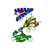
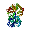
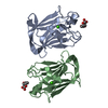
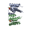
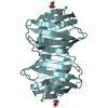
| Entry | Database: PDB / ID: 6c5d | ||||||
|---|---|---|---|---|---|---|---|
| Title | N-terminal domain of Helicobacter pylori LlaJI.R1 | ||||||
 Components Components | LlaJI.R1 | ||||||
 Keywords Keywords | DNA BINDING PROTEIN / B3 domain / restriction endonuclease | ||||||
| Function / homology | : / ATPase, dynein-related, AAA domain / AAA domain (dynein-related subfamily) / ATP hydrolysis activity / P-loop containing nucleoside triphosphate hydrolase / ATP binding / Restriction enzyme Function and homology information Function and homology information | ||||||
| Biological species |   Helicobacter pylori (bacteria) Helicobacter pylori (bacteria) | ||||||
| Method |  X-RAY DIFFRACTION / SYNCHROTRON / MOLECULAR REPLACEMENT / Resolution: 1.97 Å X-RAY DIFFRACTION / SYNCHROTRON / MOLECULAR REPLACEMENT / Resolution: 1.97 Å | ||||||
 Authors Authors | Hosford, C.J. / Chappie, J.S. | ||||||
 Citation Citation | Journal: J. Biol. Chem. / Year: 2018 Title: The crystal structure of theHelicobacter pyloriLlaJI.R1 N-terminal domain provides a model for site-specific DNA binding. Authors: Hosford, C.J. / Chappie, J.S. | ||||||
| History |
|
- Structure visualization
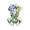
| Structure viewer | Molecule: MolmilJmol/JSmol |
|---|
- Downloads & links
-Download
| PDBx/mmCIF format | 6c5d.cif.gz | 118.4 KB | Display | PDBx/mmCIF format |
|---|---|---|---|---|
| PDB format | pdb6c5d.ent.gz | 93.2 KB | Display | PDB format |
| PDBx/mmJSON format | 6c5d.json.gz | Tree view | PDBx/mmJSON format | |
| Others |  Other downloads Other downloads |
-Validation report
| Arichive directory | https://data.pdbj.org/pub/pdb/validation_reports/c5/6c5dftp://data.pdbj.org/pub/pdb/validation_reports/c5/6c5d | HTTPS FTP |
|---|
-Related structure data
| Similar structure data |
|---|


-Links
 PDBj
PDBj- Assembly
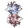
| Deposited unit | 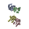
| ||||||||
|---|---|---|---|---|---|---|---|---|---|
| 1 | 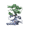
| ||||||||
| 2 | 
| ||||||||
| Unit cell |
|
-Components
| #1: Protein | Mass: 17006.238 Da / Num. of mol.: 4 / Fragment: N-terminal domain (UNP residues 1-137) Source method: isolated from a genetically manipulated source Source: (gene. exp.) Helicobacter pylori (bacteria) / Strain: J99 / ATCC 700824 / Gene: jhp_0164 / Production host: #2: Water | ChemComp-HOH / |  Mass: 18.015 Da / Num. of mol.: 245 / Source method: isolated from a natural source / Formula: H2O Mass: 18.015 Da / Num. of mol.: 245 / Source method: isolated from a natural source / Formula: H2OHas protein modification | Y | |
|---|
-Experimental details
-Experiment
| Experiment | Method: X-RAY DIFFRACTION / Number of used crystals: 1 |
|---|
- Sample preparation
| Crystal | Density Matthews: 2.01 Å3/Da / Density % sol: 38.77 % |
|---|---|
| Crystal grow | Temperature: 298 K / Method: vapor diffusion, sitting drop / pH: 6.5 / Details: 0.1 M MMT, pH 6.5, 25% v/v PEG1500 |
-Data collection
| Diffraction | Mean temperature: 100 K |
|---|---|
| Diffraction source | Source: SYNCHROTRON / Site: APS  / Beamline: 24-ID-C / Wavelength: 0.9791 Å / Beamline: 24-ID-C / Wavelength: 0.9791 Å |
| Detector | Type: DECTRIS PILATUS 6M-F / Detector: PIXEL / Date: Apr 21, 2017 |
| Radiation | Monochromator: Cryo-cooled double crystal Si(111) / Protocol: SINGLE WAVELENGTH / Monochromatic (M) / Laue (L): M / Scattering type: x-ray |
| Radiation wavelength | Wavelength: 0.9791 Å / Relative weight: 1 |
| Reflection | Resolution: 1.97→83.97 Å / Num. obs: 152937 / % possible obs: 93.6 % / Redundancy: 3.68 % / CC1/2: 0.997 / Rrim(I) all: 0.096 / Net I/σ(I): 9 |
| Reflection shell | Resolution: 1.97→2.1 Å / Redundancy: 1.91 % / Mean I/σ(I) obs: 1.56 / Num. unique obs: 23180 / CC1/2: 0.709 / Rrim(I) all: 0.624 / % possible all: 90.1 |
- Processing
| Software |
| ||||||||||||
|---|---|---|---|---|---|---|---|---|---|---|---|---|---|
| Refinement | Method to determine structure: MOLECULAR REPLACEMENT / Resolution: 1.97→83.97 Å / Cross valid method: FREE R-VALUE /
| ||||||||||||
| Refinement step | Cycle: LAST / Resolution: 1.97→83.97 Å
|
