[English] 日本語
Yorodumi
- PDB-6brd: Crystal structure of rifampin monooxygenase from Streptomyces ven... -

+
Open data


ID or keywords:

Loading...

-
Basic information

Entry
Database: PDB / ID: 6brd
Title
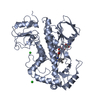
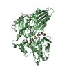
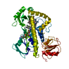
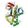
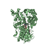
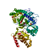
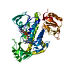
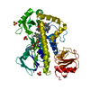
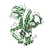
Crystal structure of rifampin monooxygenase from Streptomyces venezuelae, complexed with rifampin and FAD
ComponentsRifampin monooxygenase
KeywordsOXIDOREDUCTASE / rifampin / rifamycin / antibiotic resistance / monooxygenase / FAD / flavin adenine dinucleotide / structural genomics / CSGID / Center for Structural Genomics of Infectious Diseases / NIAID / National Institute of Allergy and Infectious Disease
Function / homology
Function and homology information


rifampicin monooxygenase / monooxygenase activity / FAD binding
Similarity search - Function
Alpha-Beta Plaits - #2450 / Glutaredoxin - #120 / FAD-binding domain / FAD binding domain / FAD/NAD(P)-binding domain / FAD/NAD(P)-binding domain / 3-Layer(bba) Sandwich / Glutaredoxin / FAD/NAD(P)-binding domain superfamily / Alpha-Beta Plaits ...Alpha-Beta Plaits - #2450 / Glutaredoxin - #120 / FAD-binding domain / FAD binding domain / FAD/NAD(P)-binding domain / FAD/NAD(P)-binding domain / 3-Layer(bba) Sandwich / Glutaredoxin / FAD/NAD(P)-binding domain superfamily / Alpha-Beta Plaits / 2-Layer Sandwich / 3-Layer(aba) Sandwich / Alpha Beta
Similarity search - Domain/homology
FLAVIN-ADENINE DINUCLEOTIDE / RIFAMPICIN / Rifampicin monooxygenase
Similarity search - Component
Biological speciesStreptomyces venezuelae (bacteria)
MethodX-RAY DIFFRACTION / MOLECULAR REPLACEMENT / Resolution: 3.32 Å
AuthorsCox, G. / Kelso, J. / Stogios, P.J. / Savchenko, A. / Anderson, W.F. / Wright, G.D. / Center for Structural Genomics of Infectious Diseases (CSGID)
Funding support United States, 1items
OrganizationGrant numberCountry
National Institutes of Health/National Institute Of Allergy and Infectious Diseases (NIH/NIAID)HHSN2720120026C United States
CitationJournal: Cell Chem Biol / Year: 2018
Title: Rox, a Rifamycin Resistance Enzyme with an Unprecedented Mechanism of Action.
Authors: Koteva, K. / Cox, G. / Kelso, J.K. / Surette, M.D. / Zubyk, H.L. / Ejim, L. / Stogios, P. / Savchenko, A. / Sorensen, D. / Wright, G.D.
History
DepositionNov 30, 2017Deposition site: RCSB / Processing site: RCSB
SupersessionDec 13, 2017ID: 5VQC
Revision 1.0Dec 13, 2017Provider: repository / Type: Initial release
Revision 1.1Jan 17, 2018Group: Author supporting evidence / Category: pdbx_audit_support / Item: _pdbx_audit_support.funding_organization
Revision 1.2Feb 21, 2018Group: Database references
Category: citation / citation_author / pdbx_database_related
Item: _citation.country / _citation.journal_abbrev ..._citation.country / _citation.journal_abbrev / _citation.journal_id_CSD / _citation.journal_id_ISSN / _citation.pdbx_database_id_DOI / _citation.pdbx_database_id_PubMed / _citation.title / _citation.year
Revision 1.3May 2, 2018Group: Data collection / Database references / Category: citation
Item: _citation.journal_volume / _citation.page_first / _citation.page_last
Revision 1.4Dec 11, 2019Group: Author supporting evidence / Category: pdbx_audit_support / Item: _pdbx_audit_support.funding_organization
Revision 1.5Oct 4, 2023Group: Data collection / Database references ...Data collection / Database references / Derived calculations / Refinement description
Category: chem_comp_atom / chem_comp_bond ...chem_comp_atom / chem_comp_bond / database_2 / pdbx_initial_refinement_model / pdbx_struct_conn_angle / struct_conn
Item: _database_2.pdbx_DOI / _database_2.pdbx_database_accession ..._database_2.pdbx_DOI / _database_2.pdbx_database_accession / _pdbx_struct_conn_angle.ptnr1_auth_asym_id / _pdbx_struct_conn_angle.ptnr1_auth_comp_id / _pdbx_struct_conn_angle.ptnr1_auth_seq_id / _pdbx_struct_conn_angle.ptnr1_label_asym_id / _pdbx_struct_conn_angle.ptnr1_label_atom_id / _pdbx_struct_conn_angle.ptnr1_label_comp_id / _pdbx_struct_conn_angle.ptnr1_label_seq_id / _pdbx_struct_conn_angle.ptnr1_symmetry / _pdbx_struct_conn_angle.ptnr3_auth_asym_id / _pdbx_struct_conn_angle.ptnr3_auth_comp_id / _pdbx_struct_conn_angle.ptnr3_auth_seq_id / _pdbx_struct_conn_angle.ptnr3_label_asym_id / _pdbx_struct_conn_angle.ptnr3_label_atom_id / _pdbx_struct_conn_angle.ptnr3_label_comp_id / _pdbx_struct_conn_angle.ptnr3_label_seq_id / _pdbx_struct_conn_angle.ptnr3_symmetry / _pdbx_struct_conn_angle.value / _struct_conn.pdbx_dist_value / _struct_conn.ptnr1_auth_asym_id / _struct_conn.ptnr1_auth_comp_id / _struct_conn.ptnr1_auth_seq_id / _struct_conn.ptnr1_label_asym_id / _struct_conn.ptnr1_label_atom_id / _struct_conn.ptnr1_label_comp_id / _struct_conn.ptnr1_label_seq_id / _struct_conn.ptnr1_symmetry / _struct_conn.ptnr2_auth_asym_id / _struct_conn.ptnr2_auth_comp_id / _struct_conn.ptnr2_auth_seq_id / _struct_conn.ptnr2_label_asym_id / _struct_conn.ptnr2_label_atom_id / _struct_conn.ptnr2_label_comp_id / _struct_conn.ptnr2_symmetry

-
Structure visualization

Structure viewerMolecule:
MolmilJmol/JSmol

Downloads & links

-
Assembly

Deposited unit
A: Rifampin monooxygenase
B: Rifampin monooxygenase
C: Rifampin monooxygenase
hetero molecules


Theoretical massNumber of molelcules
Total (without water)162,61520
Polymers157,4113
Non-polymers5,20417
Water1,63991
1
A: Rifampin monooxygenase
hetero molecules


Theoretical massNumber of molelcules
Total (without water)54,2217
Polymers52,4701
Non-polymers1,7506
Water181
TypeNameSymmetry operationNumber
identity operation1_555x,y,z1
2
B: Rifampin monooxygenase
hetero molecules


Theoretical massNumber of molelcules
Total (without water)54,2097
Polymers52,4701
Non-polymers1,7396
Water181
TypeNameSymmetry operationNumber
identity operation1_555x,y,z1
3
C: Rifampin monooxygenase
hetero molecules


Theoretical massNumber of molelcules
Total (without water)54,1856
Polymers52,4701
Non-polymers1,7155
Water181
TypeNameSymmetry operationNumber
identity operation1_555x,y,z1
Unit cell
Length a, b, c (Å)202.026, 129.402, 74.342
Angle α, β, γ (deg.)90.00, 105.45, 90.00
Int Tables number5
Space group name H-MC121
Components on special symmetry positions
IDModelComponents
11B-503-

CL

-
Components

-
Protein , 1 types, 3 molecules ABC

#1: Protein Rifampin monooxygenase / ROX


Mass: 52470.309 Da / Num. of mol.: 3
Source method: isolated from a genetically manipulated source
Source: (gene. exp.) Streptomyces venezuelae (strain ATCC 10712 / CBS 650.69 / DSM 40230 / JCM 4526 / NBRC 13096 / PD 04745) (bacteria)
Strain: ATCC 10712 / CBS 650.69 / DSM 40230 / JCM 4526 / NBRC 13096 / PD 04745
Gene: SVEN_0481 / Production host: Escherichia coli BL21(DE3) (bacteria) / Strain (production host): BL21(DE3) / References: UniProt: F2R776

-
Non-polymers , 5 types, 108 molecules

#2: Chemical ChemComp-FAD / FLAVIN-ADENINE DINUCLEOTIDE


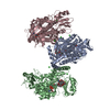
Mass: 785.550 Da / Num. of mol.: 3 / Source method: obtained synthetically / Formula: C27H33N9O15P2 / Comment: FAD*YM
#3: Chemical ChemComp-RFP / RIFAMPICIN


Mass: 822.940 Da / Num. of mol.: 3 / Source method: obtained synthetically / Formula: C43H58N4O12 / Comment: antibiotic*YM
#4: Chemical
ChemComp-CL / CHLORIDE ION


Mass: 35.453 Da / Num. of mol.: 10 / Source method: obtained synthetically / Formula: Cl
#5: Chemical ChemComp-MG / MAGNESIUM ION


Mass: 24.305 Da / Num. of mol.: 1 / Source method: obtained synthetically / Formula: Mg
#6: Water ChemComp-HOH / water


Mass: 18.015 Da / Num. of mol.: 91 / Source method: isolated from a natural source / Formula: H2O

-
Experimental details

-
Experiment

ExperimentMethod: X-RAY DIFFRACTION / Number of used crystals: 1

-
Sample preparation

CrystalDensity Matthews: 2.98 Å3/Da / Density % sol: 58.66 %
Crystal growTemperature: 298 K / Method: vapor diffusion, sitting drop / pH: 6.5
Details: 0.1 M sodium cacodylate, pH 6.5, 20% w/v PEG8000, 0.2 M magnesium acetate, soaked in rifamycin

-
Data collection

DiffractionMean temperature: 100 K
Diffraction sourceSource: ROTATING ANODE / Type: RIGAKU MICROMAX-007 / Wavelength: 1.5418 Å
DetectorType: RIGAKU RAXIS IV++ / Detector: IMAGE PLATE / Date: Apr 11, 2017
RadiationProtocol: SINGLE WAVELENGTH / Monochromatic (M) / Laue (L): M / Scattering type: x-ray
Radiation wavelengthWavelength: 1.5418 Å / Relative weight: 1
ReflectionResolution: 3.3→33.417 Å / Num. obs: 27436 / % possible obs: 97.1 % / Redundancy: 5.8 % / Rsym value: 0.162 / Net I/σ(I): 5.55
Reflection shellResolution: 3.3→3.36 Å / Redundancy: 4.3 % / Mean I/σ(I) obs: 1.33 / Rsym value: 0.443 / % possible all: 83.3

-
Processing

Software
NameVersionClassification
PHENIX(dev_2733: ???)refinement
HKL-3000data reduction
HKL-3000data scaling
PHENIXphasing
PHENIXmodel building
Cootmodel building
RefinementMethod to determine structure: MOLECULAR REPLACEMENT
Starting model: PDB entry 5KOW
Resolution: 3.32→33.417 Å / SU ML: 0.24 / Cross valid method: FREE R-VALUE / σ(F): 1.39 / Phase error: 21.63
RfactorNum. reflection% reflectionSelection details
Rfree0.2344 1333 5 %RANDOM
Rwork0.1845 ---
obs0.187 26650 97.75 %-
Solvent computationShrinkage radii: 0.9 Å / VDW probe radii: 1.11 Å
Refinement stepCycle: LAST / Resolution: 3.32→33.417 Å
ProteinNucleic acidLigandSolventTotal
Num. atoms10793 0 326 92 11211
Refine LS restraints
Refine-IDTypeDev idealNumber
X-RAY DIFFRACTIONf_bond_d0.01111337
X-RAY DIFFRACTIONf_angle_d1.07915440
X-RAY DIFFRACTIONf_dihedral_angle_d20.3084029
X-RAY DIFFRACTIONf_chiral_restr0.0561724
X-RAY DIFFRACTIONf_plane_restr0.0072009
LS refinement shell
Resolution (Å)Rfactor RfreeNum. reflection RfreeRfactor RworkNum. reflection RworkRefine-ID% reflection obs (%)
3.3201-3.43860.26171210.22672298X-RAY DIFFRACTION90
3.4386-3.57610.28811280.21982449X-RAY DIFFRACTION96
3.5761-3.73870.2681350.2052545X-RAY DIFFRACTION98
3.7387-3.93550.26391340.18592552X-RAY DIFFRACTION99
3.9355-4.18170.24111340.1722540X-RAY DIFFRACTION99
4.1817-4.50380.19321350.15442550X-RAY DIFFRACTION99
4.5038-4.95570.19241350.1632586X-RAY DIFFRACTION99
4.9557-5.66980.2531360.17622566X-RAY DIFFRACTION99
5.6698-7.13190.2461360.20882588X-RAY DIFFRACTION100
7.1319-33.41890.20961390.1782643X-RAY DIFFRACTION100
Refinement TLS params.

Method: refined / Refine-ID: X-RAY DIFFRACTION

IDL112)L122)L132)L222)L232)L332)S11 (Å °)S12 (Å °)S13 (Å °)S21 (Å °)S22 (Å °)S23 (Å °)S31 (Å °)S32 (Å °)S33 (Å °)T112)T122)T132)T222)T232)T332)Origin x (Å)Origin y (Å)Origin z (Å)
12.3698-0.29910.93142.47290.73421.8929-0.0883-0.08580.09570.4660.2722-0.57890.10940.2551-0.1010.17730.0588-0.07710.2408-0.03860.236755.1068-7.900423.8196
25.78290.9611-0.19997.629-2.47425.96690.3760.50490.95860.0111-0.3611-1.2195-0.96921.24340.00640.5148-0.13960.16210.66090.10510.705660.67958.13181.5371
30.8541-0.12680.59161.93840.03511.4924-0.1081-0.29650.16840.5247-0.00620.1503-0.0521-0.32140.08370.2060.03290.07470.3136-0.00690.225639.3785-2.66328.5914
42.4118-0.0815-0.47711.0123-0.44733.3687-0.09070.2460.1126-0.06790.04130.14030.0850.01980.04750.1895-0.0732-0.22270.20840.02950.283314.45224.6926-15.4046
51.5585-1.12220.67231.0278-1.19282.5387-0.03030.1368-0.349-0.2905-0.03370.21470.51630.01040.10070.3442-0.0336-0.11650.2137-0.0510.32722.065511.4159-8.3128
62.45010.1828-1.32932.85460.51971.7776-0.1479-0.57920.73410.350.1150.0378-0.34670.3790.03270.52820.0463-0.33360.2725-0.09280.525516.13141.91583.9694
72.9656-0.8583-0.52442.93241.36272.41780.11140.082-0.0312-0.2161-0.06720.1924-0.2387-0.1526-0.02840.12780.00150.04930.1742-0.01910.320159.925828.226745.4765
87.70482.2477-1.45075.33721.10731.4890.23231.41710.5416-0.7812-0.05410.0453-0.8271-0.2771-0.27140.86110.07420.18270.7660.21450.602270.085638.933121.2593
92.571-1.0167-0.56151.51070.86331.88740.0252-0.3386-0.48990.1888-0.0624-0.04260.16640.18770.0250.15550.01550.00250.21910.0820.450572.243517.689650.7521
Refinement TLS group
IDRefine-IDRefine TLS-IDSelection details
1X-RAY DIFFRACTION1(chain A and resid 1:171)
2X-RAY DIFFRACTION2(chain A and resid 172:260)
3X-RAY DIFFRACTION3(chain A and resid 261:474)
4X-RAY DIFFRACTION4(chain B and resid 1:171)
5X-RAY DIFFRACTION5(chain B and resid 172:372)
6X-RAY DIFFRACTION6(chain B and resid 373:474)
7X-RAY DIFFRACTION7(chain C and resid 1:175)
8X-RAY DIFFRACTION8(chain C and resid 176:256)
9X-RAY DIFFRACTION9(chain C and resid 257:474)

+
About Yorodumi

-
News

-
Feb 9, 2022. New format data for meta-information of EMDB entries

New format data for meta-information of EMDB entries

  • Version 3 of the EMDB header file is now the official format.
  • The previous official version 1.9 will be removed from the archive.

Related info.:EMDB header

External links:wwPDB to switch to version 3 of the EMDB data model

-
Aug 12, 2020. Covid-19 info

Covid-19 info

URL: https://pdbjlvh1.pdbj.org/emnavi/covid19.php

New page: Covid-19 featured information page in EM Navigator.

Related info.:Covid-19 info / Mar 5, 2020. Novel coronavirus structure data

+
Mar 5, 2020. Novel coronavirus structure data

Novel coronavirus structure data

Related info.:Yorodumi Speices / Aug 12, 2020. Covid-19 info

External links:COVID-19 featured content - PDBj / Molecule of the Month (242):Coronavirus Proteases

+
Jan 31, 2019. EMDB accession codes are about to change! (news from PDBe EMDB page)

EMDB accession codes are about to change! (news from PDBe EMDB page)

  • The allocation of 4 digits for EMDB accession codes will soon come to an end. Whilst these codes will remain in use, new EMDB accession codes will include an additional digit and will expand incrementally as the available range of codes is exhausted. The current 4-digit format prefixed with “EMD-” (i.e. EMD-XXXX) will advance to a 5-digit format (i.e. EMD-XXXXX), and so on. It is currently estimated that the 4-digit codes will be depleted around Spring 2019, at which point the 5-digit format will come into force.
  • The EM Navigator/Yorodumi systems omit the EMD- prefix.

Related info.:Q: What is EMD? / ID/Accession-code notation in Yorodumi/EM Navigator

External links:EMDB Accession Codes are Changing Soon! / Contact to PDBj

+
Jul 12, 2017. Major update of PDB

Major update of PDB

  • wwPDB released updated PDB data conforming to the new PDBx/mmCIF dictionary.
  • This is a major update changing the version number from 4 to 5, and with Remediation, in which all the entries are updated.
  • In this update, many items about electron microscopy experimental information are reorganized (e.g. em_software).
  • Now, EM Navigator and Yorodumi are based on the updated data.

External links:wwPDB Remediation / Enriched Model Files Conforming to OneDep Data Standards Now Available in the PDB FTP Archive

-
Yorodumi

Thousand views of thousand structures

  • Yorodumi is a browser for structure data from EMDB, PDB, SASBDB, etc.
  • This page is also the successor to EM Navigator detail page, and also detail information page/front-end page for Omokage search.
  • The word "yorodu" (or yorozu) is an old Japanese word meaning "ten thousand". "mi" (miru) is to see.

Related info.:EMDB / PDB / SASBDB / Comparison of 3 databanks / Yorodumi Search / Aug 31, 2016. New EM Navigator & Yorodumi / Yorodumi Papers / Jmol/JSmol / Function and homology information / Changes in new EM Navigator and Yorodumi

Read more