 Movie
Movie Controller

[English] 日本語
 Yorodumi
- PDB-6bpo: The crystal structure of the Ferric-Catecholate import receptor F... -
+ Open data

- Basic information
| Entry | Database: PDB / ID: 6bpo | ||||||
|---|---|---|---|---|---|---|---|
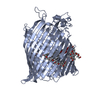
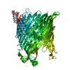
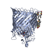
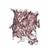
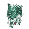
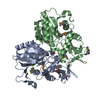
| Title | The crystal structure of the Ferric-Catecholate import receptor Fiu from K12 E. coli: Closed form (P1) | ||||||
 Components Components | Catecholate siderophore receptor Fiu | ||||||
 Keywords Keywords | MEMBRANE PROTEIN / Ferric-Catecholate / TonB-Dependent Receptor / Membrane Transport / Antibiotic Uptake / Iron Transporter | ||||||
| Function / homology |  Function and homology information Function and homology informationmicrocin transport / siderophore-iron import into cell / siderophore uptake transmembrane transporter activity / siderophore transport / cell outer membrane / signaling receptor activity / intracellular iron ion homeostasis / membrane Similarity search - Function | ||||||
| Biological species |  | ||||||
| Method |  X-RAY DIFFRACTION / SYNCHROTRON / MOLECULAR REPLACEMENT / Resolution: 2.9 Å X-RAY DIFFRACTION / SYNCHROTRON / MOLECULAR REPLACEMENT / Resolution: 2.9 Å | ||||||
 Authors Authors | Grinter, R. | ||||||
| Funding support |  United Kingdom, 1items United Kingdom, 1items
| ||||||
 Citation Citation | Journal: J.Biol.Chem. / Year: 2019 Title: The structure of the bacterial iron-catecholate transporter Fiu suggests that it imports substrates via a two-step mechanism. Authors: Grinter, R. / Lithgow, T. | ||||||
| History |
|
- Structure visualization
| Structure viewer | Molecule: MolmilJmol/JSmol |
|---|
- Downloads & links
-Download
| PDBx/mmCIF format | 6bpo.cif.gz | 1.1 MB | Display | PDBx/mmCIF format |
|---|---|---|---|---|
| PDB format | pdb6bpo.ent.gz | 900.8 KB | Display | PDB format |
| PDBx/mmJSON format | 6bpo.json.gz | Tree view | PDBx/mmJSON format | |
| Others |  Other downloads Other downloads |
-Validation report
| Arichive directory | https://data.pdbj.org/pub/pdb/validation_reports/bp/6bpoftp://data.pdbj.org/pub/pdb/validation_reports/bp/6bpo | HTTPS FTP |
|---|
-Related structure data
| Related structure data |  6bpmC  6bpnC  5fp1S S: Starting model for refinement C: citing same article ( |
|---|---|
| Similar structure data |


-Links
 PDBj
PDBj- Assembly
| Deposited unit | 
| ||||||||
|---|---|---|---|---|---|---|---|---|---|
| 1 | 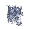
| ||||||||
| 2 | 
| ||||||||
| 3 | 
| ||||||||
| 4 | 
| ||||||||
| Unit cell |
|
-Components
| #1: Protein | Mass: 78415.461 Da / Num. of mol.: 4 Source method: isolated from a genetically manipulated source Source: (gene. exp.) Strain: K12 / Gene: fiu, ybiL, b0805, JW0790 / Production host: #2: Sugar |   Type: D-saccharide / Mass: 292.369 Da / Num. of mol.: 2 Type: D-saccharide / Mass: 292.369 Da / Num. of mol.: 2Source method: isolated from a genetically manipulated source Formula: C14H28O6 / Comment: detergent*YM #3: Water | ChemComp-HOH / |  Mass: 18.015 Da / Num. of mol.: 256 / Source method: isolated from a natural source / Formula: H2O Mass: 18.015 Da / Num. of mol.: 256 / Source method: isolated from a natural source / Formula: H2OHas protein modification | Y | |
|---|
-Experimental details
-Experiment
| Experiment | Method: X-RAY DIFFRACTION / Number of used crystals: 1 |
|---|
- Sample preparation
| Crystal | Density Matthews: 3.99 Å3/Da / Density % sol: 69.2 % |
|---|---|
| Crystal grow | Temperature: 293 K / Method: vapor diffusion, sitting drop / pH: 8.5 Details: 0.1 M Bis-Tris Propane, 20 % PEG3350, 0.2 M Na-Malonate |
-Data collection
| Diffraction | Mean temperature: 100 K |
|---|---|
| Diffraction source | Source: SYNCHROTRON / Site: Australian Synchrotron  / Beamline: MX2 / Wavelength: 0.987 Å / Beamline: MX2 / Wavelength: 0.987 Å |
| Detector | Type: DECTRIS EIGER X 16M / Detector: PIXEL / Date: Nov 6, 2017 |
| Radiation | Protocol: SINGLE WAVELENGTH / Monochromatic (M) / Laue (L): M / Scattering type: x-ray |
| Radiation wavelength | Wavelength: 0.987 Å / Relative weight: 1 |
| Reflection | Resolution: 2.9→50.31 Å / Num. obs: 116009 / % possible obs: 99 % / Observed criterion σ(I): 1.1 / Redundancy: 3.7 % / Biso Wilson estimate: 58.4 Å2 / Rmerge(I) obs: 0.222 / Rpim(I) all: 0.151 / Net I/σ(I): 3.6 |
| Reflection shell | Resolution: 2.9→2.95 Å / Redundancy: 3.7 % / Rmerge(I) obs: 1.199 / Num. unique obs: 5714 / CC1/2: 0.491 / Rpim(I) all: 0.802 / % possible all: 98.5 |
- Processing
| Software |
| |||||||||||||||||||||||||||||||||||||||||||||||||||||||||||||||||||||||||||||||||||||||||||||||||||||||||||||||||||||||||||||
|---|---|---|---|---|---|---|---|---|---|---|---|---|---|---|---|---|---|---|---|---|---|---|---|---|---|---|---|---|---|---|---|---|---|---|---|---|---|---|---|---|---|---|---|---|---|---|---|---|---|---|---|---|---|---|---|---|---|---|---|---|---|---|---|---|---|---|---|---|---|---|---|---|---|---|---|---|---|---|---|---|---|---|---|---|---|---|---|---|---|---|---|---|---|---|---|---|---|---|---|---|---|---|---|---|---|---|---|---|---|---|---|---|---|---|---|---|---|---|---|---|---|---|---|---|---|---|
| Refinement | Method to determine structure: MOLECULAR REPLACEMENT Starting model: 5FP1 Resolution: 2.9→29.23 Å / Cor.coef. Fo:Fc: 0.884 / Cor.coef. Fo:Fc free: 0.851 / SU R Cruickshank DPI: 0.509 / Cross valid method: THROUGHOUT / σ(F): 0 / SU R Blow DPI: 0.448 / SU Rfree Blow DPI: 0.287 / SU Rfree Cruickshank DPI: 0.301
| |||||||||||||||||||||||||||||||||||||||||||||||||||||||||||||||||||||||||||||||||||||||||||||||||||||||||||||||||||||||||||||
| Displacement parameters | Biso mean: 56.2 Å2
| |||||||||||||||||||||||||||||||||||||||||||||||||||||||||||||||||||||||||||||||||||||||||||||||||||||||||||||||||||||||||||||
| Refine analyze | Luzzati coordinate error obs: 0.4 Å | |||||||||||||||||||||||||||||||||||||||||||||||||||||||||||||||||||||||||||||||||||||||||||||||||||||||||||||||||||||||||||||
| Refinement step | Cycle: 1 / Resolution: 2.9→29.23 Å
| |||||||||||||||||||||||||||||||||||||||||||||||||||||||||||||||||||||||||||||||||||||||||||||||||||||||||||||||||||||||||||||
| Refine LS restraints |
| |||||||||||||||||||||||||||||||||||||||||||||||||||||||||||||||||||||||||||||||||||||||||||||||||||||||||||||||||||||||||||||
| LS refinement shell | Resolution: 2.9→2.98 Å / Total num. of bins used: 20
| |||||||||||||||||||||||||||||||||||||||||||||||||||||||||||||||||||||||||||||||||||||||||||||||||||||||||||||||||||||||||||||
| Refinement TLS params. | Method: refined / Refine-ID: X-RAY DIFFRACTION
| |||||||||||||||||||||||||||||||||||||||||||||||||||||||||||||||||||||||||||||||||||||||||||||||||||||||||||||||||||||||||||||
| Refinement TLS group |
|
