 Movie
Movie Controller

[English] 日本語
 Yorodumi
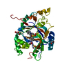
- PDB-6bh2: LINKED KDM5A JMJ DOMAIN BOUND TO THE INHIBITOR (R)-N-(1-(3-isopro... -
+ Open data

- Basic information
| Entry | Database: PDB / ID: 6bh2 | ||||||
|---|---|---|---|---|---|---|---|
| Title | LINKED KDM5A JMJ DOMAIN BOUND TO THE INHIBITOR (R)-N-(1-(3-isopropyl-1H-pyrazole-5-carbonyl)pyrrolidin-3-yl)cyclopropanecarboxamide (Compound N54) | ||||||
 Components Components | Lysine-specific demethylase 5A, linked KDM5A JMJ domain | ||||||
 Keywords Keywords | OXIDOREDUCTASE/INHIBITOR / DEMETHYLASE INHIBITION / OXIDOREDUCTASE-OXIDOREDUCTASE INHIBITOR COMPLEX / OXIDOREDUCTASE / OXIDOREDUCTASE-INHIBITOR complex | ||||||
| Function / homology |  Function and homology information Function and homology information[histone H3]-trimethyl-L-lysine4 demethylase / histone H3K4me/H3K4me2/H3K4me3 demethylase activity / facultative heterochromatin formation / histone demethylase activity / transcription initiation-coupled chromatin remodeling / enzyme inhibitor activity / Chromatin modifications during the maternal to zygotic transition (MZT) / circadian regulation of gene expression / HDMs demethylate histones / protein-DNA complex ...[histone H3]-trimethyl-L-lysine4 demethylase / histone H3K4me/H3K4me2/H3K4me3 demethylase activity / facultative heterochromatin formation / histone demethylase activity / transcription initiation-coupled chromatin remodeling / enzyme inhibitor activity / Chromatin modifications during the maternal to zygotic transition (MZT) / circadian regulation of gene expression / HDMs demethylate histones / protein-DNA complex / chromatin DNA binding / histone binding / transcription coactivator activity / transcription cis-regulatory region binding / chromatin remodeling / regulation of DNA-templated transcription / nucleolus / positive regulation of DNA-templated transcription / chromatin / negative regulation of transcription by RNA polymerase II / DNA binding / zinc ion binding / nucleoplasm / nucleus Similarity search - Function | ||||||
| Biological species |  Homo sapiens (human) Homo sapiens (human) | ||||||
| Method |  X-RAY DIFFRACTION / SYNCHROTRON / MOLECULAR REPLACEMENT / Resolution: 1.447 Å X-RAY DIFFRACTION / SYNCHROTRON / MOLECULAR REPLACEMENT / Resolution: 1.447 Å | ||||||
 Authors Authors | Horton, J.R. / Cheng, X. | ||||||
| Funding support |  United States, 1items United States, 1items
| ||||||
 Citation Citation | Journal: J. Med. Chem. / Year: 2018 Title: Insights into the Action of Inhibitor Enantiomers against Histone Lysine Demethylase 5A. Authors: Horton, J.R. / Liu, X. / Wu, L. / Zhang, K. / Shanks, J. / Zhang, X. / Rai, G. / Mott, B.T. / Jansen, D.J. / Kales, S.C. / Henderson, M.J. / Pohida, K. / Fang, Y. / Hu, X. / Jadhav, A. / ...Authors: Horton, J.R. / Liu, X. / Wu, L. / Zhang, K. / Shanks, J. / Zhang, X. / Rai, G. / Mott, B.T. / Jansen, D.J. / Kales, S.C. / Henderson, M.J. / Pohida, K. / Fang, Y. / Hu, X. / Jadhav, A. / Maloney, D.J. / Hall, M.D. / Simeonov, A. / Fu, H. / Vertino, P.M. / Yan, Q. / Cheng, X. #1: Journal: Cell Chem Biol / Year: 2016Title: Structural Basis for KDM5A Histone Lysine Demethylase Inhibition by Diverse Compounds. Authors: Horton, J.R. / Liu, X. / Gale, M. / Wu, L. / Shanks, J.R. / Zhang, X. / Webber, P.J. / Bell, J.S. / Kales, S.C. / Mott, B.T. / Rai, G. / Jansen, D.J. / Henderson, M.J. / Urban, D.J. / Hall, ...Authors: Horton, J.R. / Liu, X. / Gale, M. / Wu, L. / Shanks, J.R. / Zhang, X. / Webber, P.J. / Bell, J.S. / Kales, S.C. / Mott, B.T. / Rai, G. / Jansen, D.J. / Henderson, M.J. / Urban, D.J. / Hall, M.D. / Simeonov, A. / Maloney, D.J. / Johns, M.A. / Fu, H. / Jadhav, A. / Vertino, P.M. / Yan, Q. / Cheng, X. #2: Journal: J. Biol. Chem. / Year: 2016Title: Characterization of a Linked Jumonji Domain of the KDM5/JARID1 Family of Histone H3 Lysine 4 Demethylases. Authors: Horton, J.R. / Engstrom, A. / Zoeller, E.L. / Liu, X. / Shanks, J.R. / Zhang, X. / Johns, M.A. / Vertino, P.M. / Fu, H. / Cheng, X. | ||||||
| History |
|
- Structure visualization
| Structure viewer | Molecule: MolmilJmol/JSmol |
|---|
- Downloads & links
-Download
| PDBx/mmCIF format | 6bh2.cif.gz | 142.1 KB | Display | PDBx/mmCIF format |
|---|---|---|---|---|
| PDB format | pdb6bh2.ent.gz | 106.3 KB | Display | PDB format |
| PDBx/mmJSON format | 6bh2.json.gz | Tree view | PDBx/mmJSON format | |
| Others |  Other downloads Other downloads |
-Validation report
| Arichive directory | https://data.pdbj.org/pub/pdb/validation_reports/bh/6bh2ftp://data.pdbj.org/pub/pdb/validation_reports/bh/6bh2 | HTTPS FTP |
|---|
-Related structure data
| Related structure data |  6bguC  6bgvC  6bgwC  6bgxC  6bgyC  6bgzC  6bh0C  6bh1C  6bh3C  6bh4C  6bh5C  5e6hS S: Starting model for refinement C: citing same article ( |
|---|---|
| Similar structure data |


-Links
 PDBj
PDBj
- Assembly
| Deposited unit | 
| ||||||||
|---|---|---|---|---|---|---|---|---|---|
| 1 |
| ||||||||
| Unit cell |
|
-Components
-Protein , 1 types, 1 molecules A
| #1: Protein | Mass: 37944.836 Da / Num. of mol.: 1 Source method: isolated from a genetically manipulated source Source: (gene. exp.) Homo sapiens (human) / Gene: KDM5A, JARID1A, RBBP2, RBP2 / Production host:  References: UniProt: P29375, Oxidoreductases; Acting on paired donors, with incorporation or reduction of molecular oxygen; With 2-oxoglutarate as one donor, and incorporation of one atom of oxygen into each donor |
|---|
-Non-polymers , 5 types, 216 molecules 


| #2: Chemical | ChemComp-MN /  Mass: 54.938 Da / Num. of mol.: 1 / Source method: obtained synthetically / Formula: Mn Mass: 54.938 Da / Num. of mol.: 1 / Source method: obtained synthetically / Formula: Mn | ||||||
|---|---|---|---|---|---|---|---|
| #3: Chemical |  Mass: 92.094 Da / Num. of mol.: 2 / Source method: obtained synthetically / Formula: C3H8O3 Mass: 92.094 Da / Num. of mol.: 2 / Source method: obtained synthetically / Formula: C3H8O3#4: Chemical | ChemComp-90V / |  Mass: 290.361 Da / Num. of mol.: 1 / Source method: obtained synthetically / Formula: C15H22N4O2 / Feature type: SUBJECT OF INVESTIGATION Mass: 290.361 Da / Num. of mol.: 1 / Source method: obtained synthetically / Formula: C15H22N4O2 / Feature type: SUBJECT OF INVESTIGATION#5: Chemical | ChemComp-EDO /  Mass: 62.068 Da / Num. of mol.: 5 / Source method: obtained synthetically / Formula: C2H6O2 Mass: 62.068 Da / Num. of mol.: 5 / Source method: obtained synthetically / Formula: C2H6O2#6: Water | ChemComp-HOH / | Mass: 18.015 Da / Num. of mol.: 207 / Source method: isolated from a natural source / Formula: H2O |
-Experimental details
-Experiment
| Experiment | Method: X-RAY DIFFRACTION / Number of used crystals: 1 |
|---|
- Sample preparation
| Crystal | Density Matthews: 2.26 Å3/Da / Density % sol: 45.58 % |
|---|---|
| Crystal grow | Temperature: 289 K / Method: vapor diffusion, sitting drop Details: 1.2-1.35 M (NH4)2SO4, 0.1 M Tris-HCl (pH 8.6-9.2), 0-20% glycerol, 25 mM (Na/K) dibasic/monobasic phosphate PH range: 8.6-9.2 |
-Data collection
| Diffraction | Mean temperature: 100 K |
|---|---|
| Diffraction source | Source: SYNCHROTRON / Site: APS / Beamline: 22-ID / Wavelength: 1 Å |
| Detector | Type: RAYONIX MX300-HS / Detector: CCD / Date: Jun 17, 2016 |
| Radiation | Protocol: SINGLE WAVELENGTH / Monochromatic (M) / Laue (L): M / Scattering type: x-ray |
| Radiation wavelength | Wavelength: 1 Å / Relative weight: 1 |
| Reflection | Resolution: 1.447→33.063 Å / Num. obs: 57330 / % possible obs: 95.6 % / Redundancy: 6 % / Rmerge(I) obs: 0.11 / Rpim(I) all: 0.045 / Net I/σ(I): 16.1 |
| Reflection shell | Resolution: 1.447→1.5 Å / Redundancy: 1.6 % / Rmerge(I) obs: 0.87 / Mean I/σ(I) obs: 2 / Num. unique obs: 4064 / CC1/2: 0.389 / Rpim(I) all: 0.702 / % possible all: 68.3 |
- Processing
| Software |
| ||||||||||||||||||||||||||||||||||||||||||||||||||||||||||||||||||||||||||||||||||||||||||||||||||||||||||||||||
|---|---|---|---|---|---|---|---|---|---|---|---|---|---|---|---|---|---|---|---|---|---|---|---|---|---|---|---|---|---|---|---|---|---|---|---|---|---|---|---|---|---|---|---|---|---|---|---|---|---|---|---|---|---|---|---|---|---|---|---|---|---|---|---|---|---|---|---|---|---|---|---|---|---|---|---|---|---|---|---|---|---|---|---|---|---|---|---|---|---|---|---|---|---|---|---|---|---|---|---|---|---|---|---|---|---|---|---|---|---|---|---|---|---|
| Refinement | Method to determine structure: MOLECULAR REPLACEMENT Starting model: 5E6H Resolution: 1.447→33.063 Å / SU ML: 0.2 / Cross valid method: THROUGHOUT / σ(F): 1.33 / Phase error: 23.56
| ||||||||||||||||||||||||||||||||||||||||||||||||||||||||||||||||||||||||||||||||||||||||||||||||||||||||||||||||
| Solvent computation | Shrinkage radii: 0.9 Å / VDW probe radii: 1.11 Å | ||||||||||||||||||||||||||||||||||||||||||||||||||||||||||||||||||||||||||||||||||||||||||||||||||||||||||||||||
| Refinement step | Cycle: LAST / Resolution: 1.447→33.063 Å
| ||||||||||||||||||||||||||||||||||||||||||||||||||||||||||||||||||||||||||||||||||||||||||||||||||||||||||||||||
| Refine LS restraints |
| ||||||||||||||||||||||||||||||||||||||||||||||||||||||||||||||||||||||||||||||||||||||||||||||||||||||||||||||||
| LS refinement shell |
| ||||||||||||||||||||||||||||||||||||||||||||||||||||||||||||||||||||||||||||||||||||||||||||||||||||||||||||||||
| Refinement TLS params. | Method: refined / Refine-ID: X-RAY DIFFRACTION
| ||||||||||||||||||||||||||||||||||||||||||||||||||||||||||||||||||||||||||||||||||||||||||||||||||||||||||||||||
| Refinement TLS group |
|
