 Movie
Movie Controller


+ Open data

- Basic information
| Entry | Database: PDB / ID: 6bg8 | |||||||||
|---|---|---|---|---|---|---|---|---|---|---|
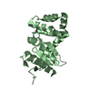
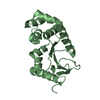
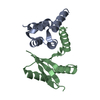
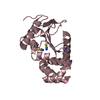
| Title | Shewanella frigidimarina ice-binding protein_1 DUF3494 Domain | |||||||||
 Components Components | Ig domain protein, group 2 domain protein | |||||||||
 Keywords Keywords | ANTIFREEZE PROTEIN / Ice Adhesin / DUF3494 Domain / C-terminal Domain | |||||||||
| Function / homology |  Function and homology information Function and homology information | |||||||||
| Biological species |  Shewanella frigidimarina (bacteria) Shewanella frigidimarina (bacteria) | |||||||||
| Method |  X-RAY DIFFRACTION / SYNCHROTRON / MOLECULAR REPLACEMENT / Resolution: 1.59712947109 Å X-RAY DIFFRACTION / SYNCHROTRON / MOLECULAR REPLACEMENT / Resolution: 1.59712947109 Å | |||||||||
 Authors Authors | Vance, T.D.R. / Davies, P.L. | |||||||||
| Funding support |  Canada, 1items Canada, 1items
| |||||||||
 Citation Citation | Journal: FEBS J. / Year: 2018 Title: An ice-binding and tandem beta-sandwich domain-containing protein in Shewanella frigidimarina is a potential new type of ice adhesin. Authors: Vance, T.D.R. / Graham, L.A. / Davies, P.L. | |||||||||
| History |
|
- Structure visualization
| Structure viewer | Molecule: MolmilJmol/JSmol |
|---|
- Downloads & links
-Download
| PDBx/mmCIF format | 6bg8.cif.gz | 148.3 KB | Display | PDBx/mmCIF format |
|---|---|---|---|---|
| PDB format | pdb6bg8.ent.gz | 92.3 KB | Display | PDB format |
| PDBx/mmJSON format | 6bg8.json.gz | Tree view | PDBx/mmJSON format | |
| Others |  Other downloads Other downloads |
-Validation report
| Arichive directory | https://data.pdbj.org/pub/pdb/validation_reports/bg/6bg8ftp://data.pdbj.org/pub/pdb/validation_reports/bg/6bg8 | HTTPS FTP |
|---|
-Related structure data
| Similar structure data |
|---|


-Links
 PDBj
PDBj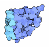


- Assembly
| Deposited unit | 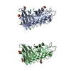
| ||||||||||||
|---|---|---|---|---|---|---|---|---|---|---|---|---|---|
| 1 | 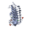
| ||||||||||||
| 2 | 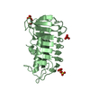
| ||||||||||||
| 3 | 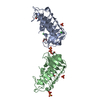
| ||||||||||||
| Unit cell |
|
-Components
-Protein , 1 types, 2 molecules AB
| #1: Protein | Mass: 25807.529 Da / Num. of mol.: 2 Source method: isolated from a genetically manipulated source Details: Density for C-terminal His tag was too unreliable to model. Source: (gene. exp.) Shewanella frigidimarina (strain NCIMB 400) (bacteria)Strain: NCIMB 400 / Gene: Sfri_1018 / Production host: |
|---|
-Non-polymers , 5 types, 787 molecules 


| #2: Chemical |  Mass: 96.063 Da / Num. of mol.: 3 / Source method: obtained synthetically / Formula: SO4 Mass: 96.063 Da / Num. of mol.: 3 / Source method: obtained synthetically / Formula: SO4#3: Chemical | ChemComp-GOL /  Mass: 92.094 Da / Num. of mol.: 4 / Source method: obtained synthetically / Formula: C3H8O3 Mass: 92.094 Da / Num. of mol.: 4 / Source method: obtained synthetically / Formula: C3H8O3#4: Chemical |  Mass: 35.453 Da / Num. of mol.: 2 / Source method: obtained synthetically / Formula: Cl Mass: 35.453 Da / Num. of mol.: 2 / Source method: obtained synthetically / Formula: Cl#5: Chemical |  Mass: 94.971 Da / Num. of mol.: 2 / Source method: obtained synthetically / Formula: PO4 Mass: 94.971 Da / Num. of mol.: 2 / Source method: obtained synthetically / Formula: PO4#6: Water | ChemComp-HOH / | Mass: 18.015 Da / Num. of mol.: 776 / Source method: isolated from a natural source / Formula: H2O |
|---|
-Experimental details
-Experiment
| Experiment | Method: X-RAY DIFFRACTION / Number of used crystals: 1 |
|---|
- Sample preparation
| Crystal | Density Matthews: 2.5 Å3/Da / Density % sol: 50.88 % |
|---|---|
| Crystal grow | Temperature: 277.15 K / Method: microbatch / Details: 2.4 M Ammonium Sulfate, 0.1 M citric acid (pH 5) |
-Data collection
| Diffraction | Mean temperature: 101.2 K |
|---|---|
| Diffraction source | Source: SYNCHROTRON / Site: CLSI / Beamline: 08B1-1 / Wavelength: 0.98 Å |
| Detector | Type: RAYONIX MX300HE / Detector: CCD / Date: May 27, 2016 |
| Radiation | Protocol: SINGLE WAVELENGTH / Monochromatic (M) / Laue (L): M / Scattering type: x-ray |
| Radiation wavelength | Wavelength: 0.98 Å / Relative weight: 1 |
| Reflection | Resolution: 1.597→42.67334 Å / Num. obs: 64562 / % possible obs: 99.14 % / Redundancy: 3.8 % / Biso Wilson estimate: 8.72197289856 Å2 / CC1/2: 0.993 / Rmerge(I) obs: 0.1775 / Net I/σ(I): 5.94 |
- Processing
| Software |
| |||||||||||||||||||||||||||||||||||||||||||||||||||||||||||||||||||||||||||||||||||||||||||||||||||||||||
|---|---|---|---|---|---|---|---|---|---|---|---|---|---|---|---|---|---|---|---|---|---|---|---|---|---|---|---|---|---|---|---|---|---|---|---|---|---|---|---|---|---|---|---|---|---|---|---|---|---|---|---|---|---|---|---|---|---|---|---|---|---|---|---|---|---|---|---|---|---|---|---|---|---|---|---|---|---|---|---|---|---|---|---|---|---|---|---|---|---|---|---|---|---|---|---|---|---|---|---|---|---|---|---|---|---|---|
| Refinement | Method to determine structure: MOLECULAR REPLACEMENT Starting model: Phyre2 Model Resolution: 1.59712947109→42.6733383803 Å / SU ML: 0.181687045676 / Cross valid method: FREE R-VALUE / σ(F): 1.34762305288 / Phase error: 25.3923917528
| |||||||||||||||||||||||||||||||||||||||||||||||||||||||||||||||||||||||||||||||||||||||||||||||||||||||||
| Solvent computation | Shrinkage radii: 0.9 Å / VDW probe radii: 1.11 Å | |||||||||||||||||||||||||||||||||||||||||||||||||||||||||||||||||||||||||||||||||||||||||||||||||||||||||
| Displacement parameters | Biso mean: 11.6712645205 Å2 | |||||||||||||||||||||||||||||||||||||||||||||||||||||||||||||||||||||||||||||||||||||||||||||||||||||||||
| Refinement step | Cycle: LAST / Resolution: 1.59712947109→42.6733383803 Å
| |||||||||||||||||||||||||||||||||||||||||||||||||||||||||||||||||||||||||||||||||||||||||||||||||||||||||
| Refine LS restraints |
| |||||||||||||||||||||||||||||||||||||||||||||||||||||||||||||||||||||||||||||||||||||||||||||||||||||||||
| LS refinement shell |
|
