| 登録情報 | データベース: PDB / ID: 6bdy
|
|---|
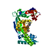
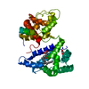
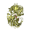
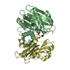
| タイトル | Crystal Structure of the MetH Reactivation Domain bound to Sinefungin |
|---|
 要素 要素 | Methionine synthase |
|---|
 キーワード キーワード | TRANSFERASE/TRANSFERASE INHIBITOR / C-terminal domain / reactivation domain / MetH-sinefungin / TRANSFERASE / TRANSFERASE-TRANSFERASE INHIBITOR complex |
|---|
| 機能・相同性 |  機能・相同性情報 機能・相同性情報
methionine synthase / methionine synthase activity / homocysteine metabolic process / methionine biosynthetic process / cobalamin binding / tetrahydrofolate metabolic process / tetrahydrofolate interconversion / methylation / zinc ion binding / cytoplasm / cytosol類似検索 - 分子機能 Cobalamin-dependent Methionine Synthase; domain 2 / Cobalamin-dependent Methionine Synthase, domain 2 / Cobalamin-dependent Methionine Synthase; domain 1 / Vitamin B12-dependent methionine synthase, activation domain / Vitamin B12-dependent methionine synthase, activation domain / Vitamin B12 dependent methionine synthase, activation domain / AdoMet activation domain profile. / Cobalamin-dependent methionine synthase / Methionine synthase, B12-binding domain / Vitamin B12-dependent methionine synthase, activation domain superfamily ...Cobalamin-dependent Methionine Synthase; domain 2 / Cobalamin-dependent Methionine Synthase, domain 2 / Cobalamin-dependent Methionine Synthase; domain 1 / Vitamin B12-dependent methionine synthase, activation domain / Vitamin B12-dependent methionine synthase, activation domain / Vitamin B12 dependent methionine synthase, activation domain / AdoMet activation domain profile. / Cobalamin-dependent methionine synthase / Methionine synthase, B12-binding domain / Vitamin B12-dependent methionine synthase, activation domain superfamily / B12-binding N-terminal domain profile. / B12 binding domain / Homocysteine-binding domain / Homocysteine-binding domain superfamily / Homocysteine S-methyltransferase / Homocysteine-binding domain profile. / : / Cobalamin (vitamin B12)-binding module, cap domain / B12 binding domain / Methionine synthase domain / B12 binding domain / Cobalamin-binding domain superfamily / B12-binding domain profile. / Cobalamin (vitamin B12)-binding domain / Pterin-binding domain / Pterin binding enzyme / Pterin-binding domain profile. / Dihydropteroate synthase-like / Roll / Orthogonal Bundle / Mainly Alpha / Alpha Beta類似検索 - ドメイン・相同性 |
|---|
| 生物種 |   Escherichia coli (大腸菌) Escherichia coli (大腸菌) |
|---|
| 手法 |  X線回折 / シンクロトロン / 分子置換 / 解像度: 1.512 Å X線回折 / シンクロトロン / 分子置換 / 解像度: 1.512 Å |
|---|
 データ登録者 データ登録者 | Fick, R.J. / Vander Lee, L.P. / Trievel, R.C. |
|---|
| 資金援助 |  米国, 1件 米国, 1件 | 組織 | 認可番号 | 国 |
|---|
| National Science Foundation (NSF, United States) | 1508492 | 米国 |
|
|---|
 引用 引用 | ジャーナル: Biochemistry / 年: 2018
タイトル: Water-Mediated Carbon-Oxygen Hydrogen Bonding Facilitates S-Adenosylmethionine Recognition in the Reactivation Domain of Cobalamin-Dependent Methionine Synthase.
著者: Fick, R.J. / Clay, M.C. / Vander Lee, L. / Scheiner, S. / Al-Hashimi, H. / Trievel, R.C. |
|---|
| 履歴 | | 登録 | 2017年10月24日 | 登録サイト: RCSB / 処理サイト: RCSB |
|---|
| 改定 1.0 | 2018年5月23日 | Provider: repository / タイプ: Initial release |
|---|
| 改定 1.1 | 2018年5月30日 | Group: Data collection / Database references / カテゴリ: citation / citation_author / Item: _citation.title / _citation_author.name |
|---|
| 改定 1.2 | 2018年7月11日 | Group: Data collection / Database references / カテゴリ: citation
Item: _citation.journal_volume / _citation.page_first / _citation.page_last |
|---|
| 改定 1.3 | 2019年11月27日 | Group: Author supporting evidence / カテゴリ: pdbx_audit_support / Item: _pdbx_audit_support.funding_organization |
|---|
| 改定 1.4 | 2023年10月4日 | Group: Data collection / Database references / Refinement description
カテゴリ: chem_comp_atom / chem_comp_bond ...chem_comp_atom / chem_comp_bond / database_2 / pdbx_initial_refinement_model
Item: _database_2.pdbx_DOI / _database_2.pdbx_database_accession |
|---|
|
|---|
 ムービー
ムービー コントローラー


 データを開く
 基本情報
基本情報 構造の表示
構造の表示 ダウンロードとリンク
 その他のダウンロード


 PDBj
PDBj
 集合体
集合体

 分子量: 381.387 Da / 分子数: 1 / 由来タイプ: 合成 / 式: C15H23N7O5
 分子量: 18.015 Da / 分子数: 442 / 由来タイプ: 天然 / 式: H2O
 試料調製
試料調製 解析
解析