 Movie
Movie Controller

[English] 日本語
 Yorodumi
- PDB-6bas: Crystal structure of Thermus thermophilus Rod shape determining p... -
+ Open data

- Basic information
| Entry | Database: PDB / ID: 6bas | ||||||
|---|---|---|---|---|---|---|---|
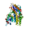
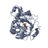
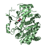
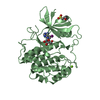
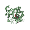
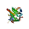
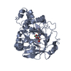
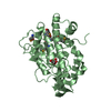
| Title | Crystal structure of Thermus thermophilus Rod shape determining protein RodA D255A mutant (Q5SIX3_THET8) | ||||||
 Components Components | Peptidoglycan glycosyltransferase RodA | ||||||
 Keywords Keywords | TRANSFERASE / Peptidoglycan Glycosyltransferase / transmembrane protein / Shape Elogation Division and Sporulation / elongasome | ||||||
| Function / homology |  Function and homology information Function and homology informationlipid-linked peptidoglycan transporter activity / peptidoglycan glycosyltransferase / peptidoglycan glycosyltransferase activity / cell division site / peptidoglycan biosynthetic process / cell wall organization / regulation of cell shape / cell division / plasma membrane Similarity search - Function | ||||||
| Biological species |   Thermus thermophilus (bacteria) Thermus thermophilus (bacteria) | ||||||
| Method |  X-RAY DIFFRACTION / SYNCHROTRON / MOLECULAR REPLACEMENT / Resolution: 3.194 Å X-RAY DIFFRACTION / SYNCHROTRON / MOLECULAR REPLACEMENT / Resolution: 3.194 Å | ||||||
 Authors Authors | Sjodt, M. / Brock, K. / Dobihal, G. / Rohs, P.D.A. / Green, A.G. / Hopf, T.A. / Meeske, A.J. / Marks, D.S. / Bernhardt, T.G. / Rudner, D.Z. / Kruse, A.C. | ||||||
| Funding support |  United States, 1items United States, 1items
| ||||||
 Citation Citation | Journal: Nature / Year: 2018 Title: Structure of the peptidoglycan polymerase RodA resolved by evolutionary coupling analysis. Authors: Sjodt, M. / Brock, K. / Dobihal, G. / Rohs, P.D.A. / Green, A.G. / Hopf, T.A. / Meeske, A.J. / Srisuknimit, V. / Kahne, D. / Walker, S. / Marks, D.S. / Bernhardt, T.G. / Rudner, D.Z. / Kruse, A.C. | ||||||
| History |
|
- Structure visualization
| Structure viewer | Molecule: MolmilJmol/JSmol |
|---|
- Downloads & links
-Download
| PDBx/mmCIF format | 6bas.cif.gz | 69.5 KB | Display | PDBx/mmCIF format |
|---|---|---|---|---|
| PDB format | pdb6bas.ent.gz | 49.8 KB | Display | PDB format |
| PDBx/mmJSON format | 6bas.json.gz | Tree view | PDBx/mmJSON format | |
| Others |  Other downloads Other downloads |
-Validation report
| Arichive directory | https://data.pdbj.org/pub/pdb/validation_reports/ba/6basftp://data.pdbj.org/pub/pdb/validation_reports/ba/6bas | HTTPS FTP |
|---|
-Related structure data
| Related structure data |  6barSC S: Starting model for refinement C: citing same article ( |
|---|---|
| Similar structure data |


-Links
 PDBj
PDBj- Assembly
| Deposited unit | 
| ||||||||
|---|---|---|---|---|---|---|---|---|---|
| 1 |
| ||||||||
| Unit cell |
|
-Components
| #1: Protein | Mass: 38644.969 Da / Num. of mol.: 1 / Mutation: D255A Source method: isolated from a genetically manipulated source Source: (gene. exp.) Thermus thermophilus (strain HB8 / ATCC 27634 / DSM 579) (bacteria)Strain: HB8 / ATCC 27634 / DSM 579 / Gene: rodA, TTHA1241 / Production host: References: UniProt: Q5SIX3, peptidoglycan glycosyltransferase |
|---|---|
| #2: Chemical | ChemComp-CL /   Mass: 35.453 Da / Num. of mol.: 1 / Source method: obtained synthetically / Formula: Cl Mass: 35.453 Da / Num. of mol.: 1 / Source method: obtained synthetically / Formula: Cl |
| #3: Water | ChemComp-HOH /  Mass: 18.015 Da / Num. of mol.: 8 / Source method: isolated from a natural source / Formula: H2O Mass: 18.015 Da / Num. of mol.: 8 / Source method: isolated from a natural source / Formula: H2O |
-Experimental details
-Experiment
| Experiment | Method: X-RAY DIFFRACTION / Number of used crystals: 1 |
|---|
- Sample preparation
| Crystal | Density Matthews: 3.03 Å3/Da / Density % sol: 59.4 % |
|---|---|
| Crystal grow | Temperature: 293 K / Method: lipidic cubic phase / pH: 8 Details: Reconstituted in 10:1 monoolein:cholesterol mix. Precipitant solution: 35-50% PEG 200, 100 mM NaCl, 100mM MgCl2, 0.1 M Tris pH 7.6-8.3 PH range: 7.6-8.3 |
-Data collection
| Diffraction | Mean temperature: 70 K |
|---|---|
| Diffraction source | Source: SYNCHROTRON / Site: APS / Beamline: 23-ID-B / Wavelength: 1.0333 Å |
| Detector | Type: DECTRIS EIGER X 16M / Detector: PIXEL / Date: Aug 18, 2017 |
| Radiation | Protocol: SINGLE WAVELENGTH / Monochromatic (M) / Laue (L): M / Scattering type: x-ray |
| Radiation wavelength | Wavelength: 1.0333 Å / Relative weight: 1 |
| Reflection | Resolution: 3.194→40.006 Å / Num. obs: 13756 / % possible obs: 99.6 % / Redundancy: 3.43 % / CC1/2: 0.996 / Rsym value: 0.171 / Net I/σ(I): 4.08 |
| Reflection shell | Resolution: 3.2→3.38 Å / Redundancy: 3.35 % / Mean I/σ(I) obs: 0.44 / Num. unique obs: 1200 / CC1/2: 0.417 / % possible all: 99.5 |
- Processing
| Software |
| |||||||||||||||||||||||||||||||||||||||||||||||||||||||||||||||||||||||||||||
|---|---|---|---|---|---|---|---|---|---|---|---|---|---|---|---|---|---|---|---|---|---|---|---|---|---|---|---|---|---|---|---|---|---|---|---|---|---|---|---|---|---|---|---|---|---|---|---|---|---|---|---|---|---|---|---|---|---|---|---|---|---|---|---|---|---|---|---|---|---|---|---|---|---|---|---|---|---|---|
| Refinement | Method to determine structure: MOLECULAR REPLACEMENT Starting model: 6BAR Resolution: 3.194→40.006 Å / SU ML: 0.64 / Cross valid method: FREE R-VALUE / σ(F): 1.34 / Phase error: 41.87 / Stereochemistry target values: ML
| |||||||||||||||||||||||||||||||||||||||||||||||||||||||||||||||||||||||||||||
| Solvent computation | Shrinkage radii: 0.9 Å / VDW probe radii: 1.11 Å / Solvent model: FLAT BULK SOLVENT MODEL | |||||||||||||||||||||||||||||||||||||||||||||||||||||||||||||||||||||||||||||
| Refinement step | Cycle: LAST / Resolution: 3.194→40.006 Å
| |||||||||||||||||||||||||||||||||||||||||||||||||||||||||||||||||||||||||||||
| Refine LS restraints |
| |||||||||||||||||||||||||||||||||||||||||||||||||||||||||||||||||||||||||||||
| LS refinement shell |
|
