 Movie
Movie Controller


+ Open data

- Basic information
| Entry | Database: PDB / ID: 6b5b | ||||||||||||
|---|---|---|---|---|---|---|---|---|---|---|---|---|---|
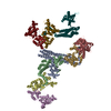
| Title | Cryo-EM structure of the NAIP5-NLRC4-flagellin inflammasome | ||||||||||||
 Components Components |
| ||||||||||||
 Keywords Keywords | IMMUNE SYSTEM / Innate immunity / molecular complex | ||||||||||||
| Function / homology |  Function and homology information Function and homology informationIPAF inflammasome complex / caspase binding / positive regulation of protein processing / cysteine-type endopeptidase inhibitor activity involved in apoptotic process / bacterial-type flagellum / pyroptotic inflammatory response / detection of bacterium / negative regulation of tumor necrosis factor-mediated signaling pathway / endopeptidase activator activity / activation of innate immune response ...IPAF inflammasome complex / caspase binding / positive regulation of protein processing / cysteine-type endopeptidase inhibitor activity involved in apoptotic process / bacterial-type flagellum / pyroptotic inflammatory response / detection of bacterium / negative regulation of tumor necrosis factor-mediated signaling pathway / endopeptidase activator activity / activation of innate immune response / positive regulation of interleukin-1 beta production / protein serine/threonine kinase binding / protein homooligomerization / positive regulation of JNK cascade / regulation of apoptotic process / defense response to Gram-negative bacterium / defense response to bacterium / positive regulation of apoptotic process / inflammatory response / innate immune response / apoptotic process / symbiont entry into host cell / negative regulation of apoptotic process / structural molecule activity / protein homodimerization activity / extracellular region / ATP binding / metal ion binding / identical protein binding / plasma membrane / cytosol Similarity search - Function | ||||||||||||
| Biological species |    Legionella pneumophila (bacteria) Legionella pneumophila (bacteria) | ||||||||||||
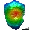
| Method | ELECTRON MICROSCOPY / single particle reconstruction / cryo EM / Resolution: 5.2 Å | ||||||||||||
 Authors Authors | Tenthorey, J.L. / Haloupek, N. / Lopez-Blanco, J.R. / Grob, P. / Adamson, E. / Hartenian, E. / Lind, N.A. / Bourgeois, N.M. / Chacon, P. / Nogales, E. / Vance, R.E. | ||||||||||||
| Funding support |  Spain, Spain,  United States, 3items United States, 3items
| ||||||||||||
 Citation Citation | Journal: Science / Year: 2017 Title: The structural basis of flagellin detection by NAIP5: A strategy to limit pathogen immune evasion. Authors: Jeannette L Tenthorey / Nicole Haloupek / José Ramón López-Blanco / Patricia Grob / Elise Adamson / Ella Hartenian / Nicholas A Lind / Natasha M Bourgeois / Pablo Chacón / Eva Nogales / Russell E Vance / Abstract: Robust innate immune detection of rapidly evolving pathogens is critical for host defense. Nucleotide-binding domain leucine-rich repeat (NLR) proteins function as cytosolic innate immune sensors in ...Robust innate immune detection of rapidly evolving pathogens is critical for host defense. Nucleotide-binding domain leucine-rich repeat (NLR) proteins function as cytosolic innate immune sensors in plants and animals. However, the structural basis for ligand-induced NLR activation has so far remained unknown. NAIP5 (NLR family, apoptosis inhibitory protein 5) binds the bacterial protein flagellin and assembles with NLRC4 to form a multiprotein complex called an inflammasome. Here we report the cryo-electron microscopy structure of the assembled ~1.4-megadalton flagellin-NAIP5-NLRC4 inflammasome, revealing how a ligand activates an NLR. Six distinct NAIP5 domains contact multiple conserved regions of flagellin, prying NAIP5 into an open and active conformation. We show that innate immune recognition of multiple ligand surfaces is a generalizable strategy that limits pathogen evolution and immune escape. | ||||||||||||
| History |
|
- Structure visualization
| Movie |
Movie viewer |
|---|---|
| Structure viewer | Molecule: MolmilJmol/JSmol |
- Downloads & links
-Download
| PDBx/mmCIF format | 6b5b.cif.gz | 562.9 KB | Display | PDBx/mmCIF format |
|---|---|---|---|---|
| PDB format | pdb6b5b.ent.gz | 447.3 KB | Display | PDB format |
| PDBx/mmJSON format | 6b5b.json.gz | Tree view | PDBx/mmJSON format | |
| Others |  Other downloads Other downloads |
-Validation report
| Arichive directory | https://data.pdbj.org/pub/pdb/validation_reports/b5/6b5bftp://data.pdbj.org/pub/pdb/validation_reports/b5/6b5b | HTTPS FTP |
|---|
-Related structure data
| Related structure data |  7055MC M: map data used to model this data C: citing same article ( |
|---|---|
| Similar structure data |


-Links
 PDBj
PDBj


- Assembly
| Deposited unit | 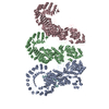
|
|---|---|
| 1 |
|
-Components
| #1: Protein | Mass: 159874.828 Da / Num. of mol.: 1 Source method: isolated from a genetically manipulated source Source: (gene. exp.)  Homo sapiens (human) / References: UniProt: Q9R016 Homo sapiens (human) / References: UniProt: Q9R016 | ||
|---|---|---|---|
| #2: Protein | Mass: 116886.000 Da / Num. of mol.: 2 Source method: isolated from a genetically manipulated source Source: (gene. exp.) Homo sapiens (human) / References: UniProt: Q3UP24#3: Protein | | Mass: 58494.070 Da / Num. of mol.: 1 Source method: isolated from a genetically manipulated source Source: (gene. exp.) Legionella pneumophila (bacteria) / Gene: fliC / Production host: Homo sapiens (human) / References: UniProt: G8UUW9, UniProt: Q5ZVV0*PLUS |
-Experimental details
-Experiment
| Experiment | Method: ELECTRON MICROSCOPY |
|---|---|
| EM experiment | Aggregation state: PARTICLE / 3D reconstruction method: single particle reconstruction |
- Sample preparation
| Component |
| |||||||||||||||||||||||||||||||||||
|---|---|---|---|---|---|---|---|---|---|---|---|---|---|---|---|---|---|---|---|---|---|---|---|---|---|---|---|---|---|---|---|---|---|---|---|---|
| Source (natural) |
| |||||||||||||||||||||||||||||||||||
| Source (recombinant) |
| |||||||||||||||||||||||||||||||||||
| Buffer solution | pH: 7.6 | |||||||||||||||||||||||||||||||||||
| Buffer component |
| |||||||||||||||||||||||||||||||||||
| Specimen | Embedding applied: NO / Shadowing applied: NO / Staining applied: NO / Vitrification applied: YES | |||||||||||||||||||||||||||||||||||
| Specimen support | Details: Carbon-coated / Grid material: COPPER / Grid mesh size: 400 divisions/in. / Grid type: Quantifoil R2/2 | |||||||||||||||||||||||||||||||||||
| Vitrification | Instrument: FEI VITROBOT MARK II / Cryogen name: ETHANE / Humidity: 100 % / Chamber temperature: 295 K |
- Electron microscopy imaging
| Experimental equipment |  Model: Titan Krios / Image courtesy: FEI Company |
|---|---|
| Microscopy | Model: FEI TITAN KRIOS |
| Electron gun | Electron source:  FIELD EMISSION GUN / Accelerating voltage: 300 kV / Illumination mode: FLOOD BEAM FIELD EMISSION GUN / Accelerating voltage: 300 kV / Illumination mode: FLOOD BEAM |
| Electron lens | Mode: BRIGHT FIELD / Nominal magnification: 22500 X / Nominal defocus max: 4000 nm / Nominal defocus min: 1800 nm |
| Image recording | Average exposure time: 6 sec. / Electron dose: 45.8 e/Å2 / Film or detector model: GATAN K2 SUMMIT (4k x 4k) / Num. of grids imaged: 3 |
| Image scans | Movie frames/image: 20 / Used frames/image: 1-20 |
- Processing
| Software | Name: PHENIX / Version: 1.11.1_2575: / Classification: refinement | |||||||||||||||||||||||||||||||||||||||||||||||||||||||||||||||||
|---|---|---|---|---|---|---|---|---|---|---|---|---|---|---|---|---|---|---|---|---|---|---|---|---|---|---|---|---|---|---|---|---|---|---|---|---|---|---|---|---|---|---|---|---|---|---|---|---|---|---|---|---|---|---|---|---|---|---|---|---|---|---|---|---|---|---|
| EM software |
| |||||||||||||||||||||||||||||||||||||||||||||||||||||||||||||||||
| CTF correction | Type: PHASE FLIPPING AND AMPLITUDE CORRECTION | |||||||||||||||||||||||||||||||||||||||||||||||||||||||||||||||||
| Particle selection | Num. of particles selected: 856358 | |||||||||||||||||||||||||||||||||||||||||||||||||||||||||||||||||
| Symmetry | Point symmetry: C1 (asymmetric) | |||||||||||||||||||||||||||||||||||||||||||||||||||||||||||||||||
| 3D reconstruction | Resolution: 5.2 Å / Resolution method: FSC 0.143 CUT-OFF / Num. of particles: 252214 / Symmetry type: POINT | |||||||||||||||||||||||||||||||||||||||||||||||||||||||||||||||||
| Atomic model building | B value: 167 / Protocol: FLEXIBLE FIT / Space: REAL / Target criteria: Correlation coefficient Details: Homology models predicted by I-TASSER server were used as initial model sources. The main structural template identified by I-TASSER for NAIP5 was the crystal structure of NLRC4 in the ...Details: Homology models predicted by I-TASSER server were used as initial model sources. The main structural template identified by I-TASSER for NAIP5 was the crystal structure of NLRC4 in the inactive conformation (PDB ID: 4KXF) that covered all the domains except the N-terminal BIR region, where homology models from several BIR domains were recognized (PDB IDs: 1SE0, 2VM5, and 1OXQ for BIR1, BIR2, and BIR3, respectively). Predictions were first rigid body docked into the density map using Chimera and/or ADP_EM. Then, the docked models were flexibly fitted with iMODFIT, if necessary. Finally, all models were refined in Phenix. | |||||||||||||||||||||||||||||||||||||||||||||||||||||||||||||||||
| Refine LS restraints |
|
