[English] 日本語
Yorodumi
- PDB-6b03: The crystal structure of the ferredoxin protease FusC in complex ... -

+
Open data


ID or keywords:

Loading...

-
Basic information

Entry
Database: PDB / ID: 6b03
TitleThe crystal structure of the ferredoxin protease FusC in complex with its substrate plant ferredoxin
Components
  • Ferredoxin-2, chloroplastic
  • Putative zinc protease
KeywordsHYDROLASE / M16 Protease / Ferredoxin cleavage / Iron Acquisition
Function / homology
Function and homology information


photosynthetic acclimation / photosynthetic electron transport chain / chloroplast / metalloendopeptidase activity / 2 iron, 2 sulfur cluster binding / electron transfer activity / proteolysis / metal ion binding
Similarity search - Function
Ferredoxin [2Fe-2S], plant / 2Fe-2S ferredoxin, iron-sulphur binding site / 2Fe-2S ferredoxin-type iron-sulfur binding region signature. / Peptidase M16, zinc-binding site / Insulinase family, zinc-binding region signature. / 2Fe-2S iron-sulfur cluster binding domain / Peptidase M16, C-terminal / Peptidase M16 inactive domain / Peptidase M16, N-terminal / Insulinase (Peptidase family M16) ...Ferredoxin [2Fe-2S], plant / 2Fe-2S ferredoxin, iron-sulphur binding site / 2Fe-2S ferredoxin-type iron-sulfur binding region signature. / Peptidase M16, zinc-binding site / Insulinase family, zinc-binding region signature. / 2Fe-2S iron-sulfur cluster binding domain / Peptidase M16, C-terminal / Peptidase M16 inactive domain / Peptidase M16, N-terminal / Insulinase (Peptidase family M16) / Metalloenzyme, LuxS/M16 peptidase-like / Beta-grasp domain superfamily / 2Fe-2S ferredoxin-type iron-sulfur binding domain profile. / 2Fe-2S ferredoxin-type iron-sulfur binding domain / 2Fe-2S ferredoxin-like superfamily
Similarity search - Domain/homology
Ferredoxin-2, chloroplastic / Zinc protease
Similarity search - Component
Biological speciesPectobacterium atrosepticum SCRI1043 (bacteria)
Arabidopsis thaliana (thale cress)
MethodX-RAY DIFFRACTION / SYNCHROTRON / Resolution: 2.7 Å
AuthorsGrinter, R.
Funding support United Kingdom, 1items
OrganizationGrant numberCountry
Wellcome Trust106077/Z/14/Z United Kingdom
Citation
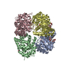
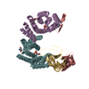
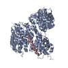
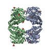
Journal: PLoS Biol / Year: 2018
Title: FusC, a member of the M16 protease family acquired by bacteria for iron piracy against plants.
Authors: Rhys Grinter / Iain D Hay / Jiangning Song / Jiawei Wang / Don Teng / Vijay Dhanesakaran / Jonathan J Wilksch / Mark R Davies / Dene Littler / Simone A Beckham / Ian R Henderson / Richard A ...Authors: Rhys Grinter / Iain D Hay / Jiangning Song / Jiawei Wang / Don Teng / Vijay Dhanesakaran / Jonathan J Wilksch / Mark R Davies / Dene Littler / Simone A Beckham / Ian R Henderson / Richard A Strugnell / Gordon Dougan / Trevor Lithgow /
Abstract: Iron is essential for life. Accessing iron from the environment can be a limiting factor that determines success in a given environmental niche. For bacteria, access of chelated iron from the ...Iron is essential for life. Accessing iron from the environment can be a limiting factor that determines success in a given environmental niche. For bacteria, access of chelated iron from the environment is often mediated by TonB-dependent transporters (TBDTs), which are β-barrel proteins that form sophisticated channels in the outer membrane. Reports of iron-bearing proteins being used as a source of iron indicate specific protein import reactions across the bacterial outer membrane. The molecular mechanism by which a folded protein can be imported in this way had remained mysterious, as did the evolutionary process that could lead to such a protein import pathway. How does the bacterium evolve the specificity factors that would be required to select and import a protein encoded on another organism's genome? We describe here a model whereby the plant iron-bearing protein ferredoxin can be imported across the outer membrane of the plant pathogen Pectobacterium by means of a Brownian ratchet mechanism, thereby liberating iron into the bacterium to enable its growth in plant tissues. This import pathway is facilitated by FusC, a member of the same protein family as the mitochondrial processing peptidase (MPP). The Brownian ratchet depends on binding sites discovered in crystal structures of FusC that engage a linear segment of the plant protein ferredoxin. Sequence relationships suggest that the bacterial gene encoding FusC has previously unappreciated homologues in plants and that the protein import mechanism employed by the bacterium is an evolutionary echo of the protein import pathway in plant mitochondria and plastids.
History
DepositionSep 13, 2017Deposition site: RCSB / Processing site: RCSB
Revision 1.0Jun 20, 2018Provider: repository / Type: Initial release
Revision 1.1Aug 15, 2018Group: Data collection / Database references / Source and taxonomy
Category: citation / citation_author / entity_src_gen
Item: _citation.country / _citation.journal_abbrev ..._citation.country / _citation.journal_abbrev / _citation.journal_id_CSD / _citation.journal_id_ISSN / _citation.journal_volume / _citation.page_first / _citation.page_last / _citation.pdbx_database_id_DOI / _citation.pdbx_database_id_PubMed / _citation.title / _citation.year / _entity_src_gen.pdbx_host_org_scientific_name
Revision 1.2Mar 13, 2024Group: Data collection / Database references / Category: chem_comp_atom / chem_comp_bond / database_2
Item: _database_2.pdbx_DOI / _database_2.pdbx_database_accession

-
Structure visualization

Structure viewerMolecule:
MolmilJmol/JSmol

Downloads & links

-
Assembly

Deposited unit
A: Putative zinc protease
F: Ferredoxin-2, chloroplastic
C: Ferredoxin-2, chloroplastic


Theoretical massNumber of molelcules
Total (without water)126,7143
Polymers126,7143
Non-polymers00
Water90150
1


  • Idetical with deposited unit
  • defined by author
  • Evidence: SAXS, isothermal titration calorimetry
TypeNameSymmetry operationNumber
identity operation1_555x,y,z1
Unit cell
Length a, b, c (Å)82.578, 127.260, 133.205
Angle α, β, γ (deg.)90.00, 90.00, 90.00
Int Tables number18
Space group name H-MP22121
Components on special symmetry positions
IDModelComponents
11A-1039-

HOH

-
Components

#1: Protein Putative zinc protease


Mass: 104028.852 Da / Num. of mol.: 1
Source method: isolated from a genetically manipulated source
Source: (gene. exp.) Pectobacterium atrosepticum SCRI1043 (bacteria)
Strain: SCRI 1043 / ATCC BAA-672 / Gene: ECA0879 / Production host: Escherichia coli BL21(DE3) (bacteria) / Strain (production host): BL21 (DE3) / Variant (production host): C41 / References: UniProt: Q6D8U3
#2: Protein Ferredoxin-2, chloroplastic / AtFd2


Mass: 11342.354 Da / Num. of mol.: 2 / Fragment: UNP residues 53-145
Source method: isolated from a genetically manipulated source
Source: (gene. exp.) Arabidopsis thaliana (thale cress) / Gene: FD2, PETF, PETF1, At1g60950, T7P1.9 / Production host: Escherichia coli BL21(DE3) (bacteria) / Strain (production host): BL21 (DE3) / Variant (production host): C41 / References: UniProt: P16972
#3: Water ChemComp-HOH / water


Mass: 18.015 Da / Num. of mol.: 50 / Source method: isolated from a natural source / Formula: H2O

-
Experimental details

-
Experiment

ExperimentMethod: X-RAY DIFFRACTION / Number of used crystals: 1

-
Sample preparation

CrystalDensity Matthews: 2.76 Å3/Da / Density % sol: 55.4 % / Description: Spare Plates
Crystal growTemperature: 294 K / Method: vapor diffusion, sitting drop / pH: 7.5 / Details: 20% PEG3350, 0.2 M NaKPO4, 0.1 M BisTrisPropane

-
Data collection

DiffractionMean temperature: 100 K
Diffraction sourceSource: SYNCHROTRON / Site: Australian Synchrotron / Beamline: MX2 / Wavelength: 0.987 Å
DetectorType: MAR CCD 130 mm / Detector: CCD / Date: Feb 16, 2017 / Details: Yes
RadiationProtocol: SINGLE WAVELENGTH / Monochromatic (M) / Laue (L): M / Scattering type: x-ray
Radiation wavelengthWavelength: 0.987 Å / Relative weight: 1
ReflectionResolution: 2.7→48.01 Å / Num. obs: 39305 / % possible obs: 100 % / Observed criterion σ(I): 1.2 / Redundancy: 6.6 % / CC1/2: 0.995 / Rmerge(I) obs: 0.149 / Rpim(I) all: 0.094 / Net I/σ(I): 7.2
Reflection shellResolution: 2.7→2.81 Å / Redundancy: 6.9 % / Rmerge(I) obs: 1.363 / Mean I/σ(I) obs: 1.2 / Num. unique obs: 4382 / CC1/2: 0.538 / Rpim(I) all: 0.614 / % possible all: 100

-
Processing

Software
NameVersionClassification
PHENIX(1.11.1_2575: ???)refinement
XDSdata reduction
Aimlessdata scaling
PHASERphasing
RefinementResolution: 2.7→41.289 Å / SU ML: 0.39 / Cross valid method: FREE R-VALUE / σ(F): 1.34 / Phase error: 28.36 / Stereochemistry target values: ML
RfactorNum. reflection% reflection
Rfree0.2427 2001 5.1 %
Rwork0.1957 --
obs0.1981 39245 99.92 %
Solvent computationShrinkage radii: 0.9 Å / VDW probe radii: 1.11 Å / Solvent model: FLAT BULK SOLVENT MODEL
Refinement stepCycle: LAST / Resolution: 2.7→41.289 Å
ProteinNucleic acidLigandSolventTotal
Num. atoms7475 0 0 50 7525
Refine LS restraints
Refine-IDTypeDev idealNumber
X-RAY DIFFRACTIONf_bond_d0.0097739
X-RAY DIFFRACTIONf_angle_d1.1210515
X-RAY DIFFRACTIONf_dihedral_angle_d6.9726510
X-RAY DIFFRACTIONf_chiral_restr0.061168
X-RAY DIFFRACTIONf_plane_restr0.0071381
LS refinement shell
Resolution (Å)Rfactor RfreeNum. reflection RfreeRfactor RworkNum. reflection RworkRefine-ID% reflection obs (%)
2.7001-2.76760.42411130.342643X-RAY DIFFRACTION100
2.7676-2.84240.33921370.28862643X-RAY DIFFRACTION100
2.8424-2.9260.32581320.28592600X-RAY DIFFRACTION100
2.926-3.02040.3381450.25632654X-RAY DIFFRACTION100
3.0204-3.12830.31751610.25762581X-RAY DIFFRACTION100
3.1283-3.25360.3271450.25382641X-RAY DIFFRACTION100
3.2536-3.40160.33481310.24112643X-RAY DIFFRACTION100
3.4016-3.58080.2671390.20142639X-RAY DIFFRACTION100
3.5808-3.8050.24081430.18342648X-RAY DIFFRACTION100
3.805-4.09860.21291500.16972663X-RAY DIFFRACTION100
4.0986-4.51060.2131440.15452655X-RAY DIFFRACTION100
4.5106-5.16220.19821560.15452685X-RAY DIFFRACTION100
5.1622-6.49970.24331420.18542721X-RAY DIFFRACTION100
6.4997-41.29380.18661630.17532828X-RAY DIFFRACTION100
Refinement TLS params.Method: refined / Origin x: 10.4002 Å / Origin y: 163.3148 Å / Origin z: 17.502 Å
111213212223313233
T0.3325 Å2-0.0051 Å20.0205 Å2-0.3234 Å20.066 Å2--0.3486 Å2
L0.18 °2-0.3125 °20.1571 °2-0.1087 °2-0.0543 °2--0.0988 °2
S0.0204 Å °-0.0114 Å °-0.0771 Å °0.0239 Å °-0.0477 Å °0.0714 Å °0.0454 Å °0.0277 Å °-0.0001 Å °
Refinement TLS groupSelection details: all

+
About Yorodumi

-
News

-
Feb 9, 2022. New format data for meta-information of EMDB entries

New format data for meta-information of EMDB entries

  • Version 3 of the EMDB header file is now the official format.
  • The previous official version 1.9 will be removed from the archive.

Related info.:EMDB header

External links:wwPDB to switch to version 3 of the EMDB data model

-
Aug 12, 2020. Covid-19 info

Covid-19 info

URL: https://pdbjlvh1.pdbj.org/emnavi/covid19.php

New page: Covid-19 featured information page in EM Navigator.

Related info.:Covid-19 info / Mar 5, 2020. Novel coronavirus structure data

+
Mar 5, 2020. Novel coronavirus structure data

Novel coronavirus structure data

Related info.:Yorodumi Speices / Aug 12, 2020. Covid-19 info

External links:COVID-19 featured content - PDBj / Molecule of the Month (242):Coronavirus Proteases

+
Jan 31, 2019. EMDB accession codes are about to change! (news from PDBe EMDB page)

EMDB accession codes are about to change! (news from PDBe EMDB page)

  • The allocation of 4 digits for EMDB accession codes will soon come to an end. Whilst these codes will remain in use, new EMDB accession codes will include an additional digit and will expand incrementally as the available range of codes is exhausted. The current 4-digit format prefixed with “EMD-” (i.e. EMD-XXXX) will advance to a 5-digit format (i.e. EMD-XXXXX), and so on. It is currently estimated that the 4-digit codes will be depleted around Spring 2019, at which point the 5-digit format will come into force.
  • The EM Navigator/Yorodumi systems omit the EMD- prefix.

Related info.:Q: What is EMD? / ID/Accession-code notation in Yorodumi/EM Navigator

External links:EMDB Accession Codes are Changing Soon! / Contact to PDBj

+
Jul 12, 2017. Major update of PDB

Major update of PDB

  • wwPDB released updated PDB data conforming to the new PDBx/mmCIF dictionary.
  • This is a major update changing the version number from 4 to 5, and with Remediation, in which all the entries are updated.
  • In this update, many items about electron microscopy experimental information are reorganized (e.g. em_software).
  • Now, EM Navigator and Yorodumi are based on the updated data.

External links:wwPDB Remediation / Enriched Model Files Conforming to OneDep Data Standards Now Available in the PDB FTP Archive

-
Yorodumi

Thousand views of thousand structures

  • Yorodumi is a browser for structure data from EMDB, PDB, SASBDB, etc.
  • This page is also the successor to EM Navigator detail page, and also detail information page/front-end page for Omokage search.
  • The word "yorodu" (or yorozu) is an old Japanese word meaning "ten thousand". "mi" (miru) is to see.

Related info.:EMDB / PDB / SASBDB / Comparison of 3 databanks / Yorodumi Search / Aug 31, 2016. New EM Navigator & Yorodumi / Yorodumi Papers / Jmol/JSmol / Function and homology information / Changes in new EM Navigator and Yorodumi

Read more