 Movie
Movie Controller

[English] 日本語
 Yorodumi
- PDB-6aty: Exploring Cystine Dense Peptide Space to Open a Unique Molecular ... -
+ Open data

- Basic information
| Entry | Database: PDB / ID: 6aty | ||||||
|---|---|---|---|---|---|---|---|
| Title | Exploring Cystine Dense Peptide Space to Open a Unique Molecular Toolbox | ||||||
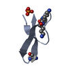
 Components Components | Venom protein 51.1 | ||||||
 Keywords Keywords | TOXIN / Knottins / Cystine knot / Toxins | ||||||
| Function / homology | toxin activity / extracellular region / Venom protein 51.1 Function and homology information Function and homology information | ||||||
| Biological species |  Lychas mucronatus (Chinese swimming scorpion) Lychas mucronatus (Chinese swimming scorpion) | ||||||
| Method |  X-RAY DIFFRACTION / SAD / Resolution: 1.8 Å X-RAY DIFFRACTION / SAD / Resolution: 1.8 Å | ||||||
 Authors Authors | Gewe, M.M. / Rupert, P. / Strong, R.K. | ||||||
 Citation Citation | Journal: To Be Published Title: Exploring Cystine Dense Peptide Space to Open a Unique Molecular Toolbox Authors: Correnti, C. / Gewe, M.M. | ||||||
| History |
|
- Structure visualization
| Structure viewer | Molecule: MolmilJmol/JSmol |
|---|
- Downloads & links
-Download
| PDBx/mmCIF format | 6aty.cif.gz | 21.6 KB | Display | PDBx/mmCIF format |
|---|---|---|---|---|
| PDB format | pdb6aty.ent.gz | 12.2 KB | Display | PDB format |
| PDBx/mmJSON format | 6aty.json.gz | Tree view | PDBx/mmJSON format | |
| Others |  Other downloads Other downloads |
-Validation report
| Arichive directory | https://data.pdbj.org/pub/pdb/validation_reports/at/6atyftp://data.pdbj.org/pub/pdb/validation_reports/at/6aty | HTTPS FTP |
|---|
-Related structure data


-Links
 PDBj
PDBj- Assembly
| Deposited unit | 
| ||||||||
|---|---|---|---|---|---|---|---|---|---|
| 1 |
| ||||||||
| 2 | x 6
| ||||||||
| 3 | 
| ||||||||
| Unit cell |
|
-Components
| #1: Protein/peptide | Mass: 4151.752 Da / Num. of mol.: 1 Source method: isolated from a genetically manipulated source Source: (gene. exp.) Lychas mucronatus (Chinese swimming scorpion)Cell line (production host): HEK-293F / Production host:  Homo sapiens (human) / References: UniProt: P0CJ17 Homo sapiens (human) / References: UniProt: P0CJ17 |
|---|---|
| #2: Chemical | ChemComp-GOL /   Mass: 92.094 Da / Num. of mol.: 1 / Source method: obtained synthetically / Formula: C3H8O3 Mass: 92.094 Da / Num. of mol.: 1 / Source method: obtained synthetically / Formula: C3H8O3 |
| #3: Water | ChemComp-HOH /  Mass: 18.015 Da / Num. of mol.: 38 / Source method: isolated from a natural source / Formula: H2O Mass: 18.015 Da / Num. of mol.: 38 / Source method: isolated from a natural source / Formula: H2O |
| Has protein modification | Y |
-Experimental details
-Experiment
| Experiment | Method: X-RAY DIFFRACTION / Number of used crystals: 1 |
|---|
- Sample preparation
| Crystal | Density Matthews: 1.89 Å3/Da / Density % sol: 34.77 % |
|---|---|
| Crystal grow | Temperature: 298 K / Method: vapor diffusion, sitting drop / pH: 4.2 Details: 0.1 M Phosphate-citrate pH 4.2, 40% Ethanol, 5% PEG 000 |
-Data collection
| Diffraction | Mean temperature: 100 K |
|---|---|
| Diffraction source | Source: ROTATING ANODE / Type: RIGAKU MICROMAX-007 HF / Wavelength: 1.54 Å |
| Detector | Type: RIGAKU SATURN 944+ / Detector: CCD / Date: Apr 8, 2016 |
| Radiation | Protocol: SINGLE WAVELENGTH / Monochromatic (M) / Laue (L): M / Scattering type: x-ray |
| Radiation wavelength | Wavelength: 1.54 Å / Relative weight: 1 |
| Reflection | Resolution: 1.8→50 Å / Num. obs: 10167 / % possible obs: 90 % / Redundancy: 16.9 % / Net I/σ(I): 105.8 |
| Reflection shell | Resolution: 1.8→1.83 Å / Redundancy: 2.3 % / Rmerge(I) obs: 0.103 / Mean I/σ(I) obs: 11.6 / Num. unique obs: 147 / CC1/2: 0.983 / Rpim(I) all: 0.67 / % possible all: 27.6 |
- Processing
| Software |
| ||||||||||||||||||||||||||||||||||||||||||||||||||||||||||||||||||||||||||||||||||||||||||||||||||||||||||||||||||||||||||||||||||||||||||||||||||||||||||||||||||||||||||||||||||||||
|---|---|---|---|---|---|---|---|---|---|---|---|---|---|---|---|---|---|---|---|---|---|---|---|---|---|---|---|---|---|---|---|---|---|---|---|---|---|---|---|---|---|---|---|---|---|---|---|---|---|---|---|---|---|---|---|---|---|---|---|---|---|---|---|---|---|---|---|---|---|---|---|---|---|---|---|---|---|---|---|---|---|---|---|---|---|---|---|---|---|---|---|---|---|---|---|---|---|---|---|---|---|---|---|---|---|---|---|---|---|---|---|---|---|---|---|---|---|---|---|---|---|---|---|---|---|---|---|---|---|---|---|---|---|---|---|---|---|---|---|---|---|---|---|---|---|---|---|---|---|---|---|---|---|---|---|---|---|---|---|---|---|---|---|---|---|---|---|---|---|---|---|---|---|---|---|---|---|---|---|---|---|---|---|
| Refinement | Method to determine structure: SAD / Resolution: 1.8→34.45 Å / Cor.coef. Fo:Fc: 0.963 / Cor.coef. Fo:Fc free: 0.937 / SU B: 2.996 / SU ML: 0.094 / Cross valid method: THROUGHOUT / ESU R: 0.143 / ESU R Free: 0.142 / Details: HYDROGENS HAVE BEEN ADDED IN THE RIDING POSITIONS
| ||||||||||||||||||||||||||||||||||||||||||||||||||||||||||||||||||||||||||||||||||||||||||||||||||||||||||||||||||||||||||||||||||||||||||||||||||||||||||||||||||||||||||||||||||||||
| Solvent computation | Ion probe radii: 0.8 Å / Shrinkage radii: 0.8 Å / VDW probe radii: 1.2 Å | ||||||||||||||||||||||||||||||||||||||||||||||||||||||||||||||||||||||||||||||||||||||||||||||||||||||||||||||||||||||||||||||||||||||||||||||||||||||||||||||||||||||||||||||||||||||
| Displacement parameters | Biso mean: 18.233 Å2
| ||||||||||||||||||||||||||||||||||||||||||||||||||||||||||||||||||||||||||||||||||||||||||||||||||||||||||||||||||||||||||||||||||||||||||||||||||||||||||||||||||||||||||||||||||||||
| Refinement step | Cycle: 1 / Resolution: 1.8→34.45 Å
| ||||||||||||||||||||||||||||||||||||||||||||||||||||||||||||||||||||||||||||||||||||||||||||||||||||||||||||||||||||||||||||||||||||||||||||||||||||||||||||||||||||||||||||||||||||||
| Refine LS restraints |
|