[English] 日本語
Yorodumi
- PDB-3hjd: X-ray structure of monomeric variant of HNP1 -

+
Open data


ID or keywords:

Loading...

-
Basic information

Entry
Database: PDB / ID: 3hjd
TitleX-ray structure of monomeric variant of HNP1
ComponentsHuman neutrophil peptide 1
KeywordsANTIMICROBIAL PROTEIN / HNP1 / monomeric defensin / antimicrobial / chemotactic / Antibiotic / Antiviral defense / Fungicide
Function / homology
Function and homology information


pore-forming activity / Defensins / disruption of plasma membrane integrity in another organism / killing by host of symbiont cells / T cell chemotaxis / Alpha-defensins / defense response to protozoan / estrogen receptor signaling pathway / defense response to fungus / innate immune response in mucosa ...pore-forming activity / Defensins / disruption of plasma membrane integrity in another organism / killing by host of symbiont cells / T cell chemotaxis / Alpha-defensins / defense response to protozoan / estrogen receptor signaling pathway / defense response to fungus / innate immune response in mucosa / Golgi lumen / antimicrobial humoral immune response mediated by antimicrobial peptide / chemotaxis / azurophil granule lumen / antibacterial humoral response / cellular response to lipopolysaccharide / collagen-containing extracellular matrix / defense response to Gram-negative bacterium / defense response to virus / killing of cells of another organism / defense response to Gram-positive bacterium / immune response / Neutrophil degranulation / extracellular space / extracellular exosome / extracellular region
Similarity search - Function
Mammalian defensins signature. / Alpha-defensin, C-terminal / Mammalian defensin / Defensin propeptide / Alpha-defensin propeptide / Alpha-defensin / Defensin propeptide / Beta/alpha-defensin, C-terminal / Defensin/corticostatin family
Similarity search - Domain/homology
Neutrophil defensin 1
Similarity search - Component
Biological speciesHomo sapiens (human)
MethodX-RAY DIFFRACTION / SYNCHROTRON / MOLECULAR REPLACEMENT / Resolution: 1.65 Å
AuthorsLubkowski, J. / Pazgier, M. / Lu, W.
CitationJournal: To be Published
Title: What Dictates the Multifaced Functions of the Human alpha-Defensin HNP?
Authors: Wei, G. / de Leeuw, E. / Pazgier, M. / Rajabi, M. / Li, J. / Zou, G. / Ericksen, B. / Wu, Z. / Yuan, W. / Szmacinski, H. / Lu, W.-Y. / Lubkowski, J. / Lehrer, R.L. / Lu, W.
History
DepositionMay 21, 2009Deposition site: RCSB / Processing site: RCSB
Revision 1.0Oct 13, 2009Provider: repository / Type: Initial release
Revision 1.1Jul 13, 2011Group: Version format compliance
Revision 1.2Sep 7, 2011Group: Advisory
Revision 1.3Jan 24, 2018Group: Database references / Category: citation_author / Item: _citation_author.name
Revision 1.4Sep 6, 2023Group: Data collection / Database references ...Data collection / Database references / Derived calculations / Refinement description
Category: chem_comp_atom / chem_comp_bond ...chem_comp_atom / chem_comp_bond / database_2 / pdbx_initial_refinement_model / struct_conn
Item: _database_2.pdbx_DOI / _database_2.pdbx_database_accession / _struct_conn.pdbx_leaving_atom_flag

-
Structure visualization

Structure viewerMolecule:
MolmilJmol/JSmol

Downloads & links

-
Assembly

Deposited unit
A: Human neutrophil peptide 1
B: Human neutrophil peptide 1


Theoretical massNumber of molelcules
Total (without water)6,9322
Polymers6,9322
Non-polymers00
Water1,31573
1
A: Human neutrophil peptide 1


Theoretical massNumber of molelcules
Total (without water)3,4661
Polymers3,4661
Non-polymers00
Water181
TypeNameSymmetry operationNumber
identity operation1_555x,y,z1
MethodPISA
2
B: Human neutrophil peptide 1


Theoretical massNumber of molelcules
Total (without water)3,4661
Polymers3,4661
Non-polymers00
Water181
TypeNameSymmetry operationNumber
identity operation1_555x,y,z1
MethodPISA
Unit cell
Length a, b, c (Å)69.85, 69.85, 46.33
Angle α, β, γ (deg.)90, 90, 120
Int Tables number177
Space group name H-MP622
Components on special symmetry positions
IDModelComponents
11A-231-

HOH

Details
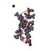
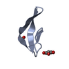
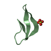
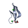
Each of two monomers present in the asymmetric unit represents half of the biological assembly. Biologically-relevant dimers are not present in crystals of this derivative

-
Components

#1: Protein/peptide Human neutrophil peptide 1 / Neutrophil defensin 1 / HNP-1 / HP-1 / HP1 / Defensin / alpha 1 / HP 1-56 / Neutrophil defensin 2 / ...Neutrophil defensin 1 / HNP-1 / HP-1 / HP1 / Defensin / alpha 1 / HP 1-56 / Neutrophil defensin 2 / HNP-2 / HP-2 / HP2


Mass: 3466.137 Da / Num. of mol.: 2 / Fragment: Residues 65-94 / Mutation: ILE84IML / Source method: obtained synthetically / Source: (synth.) Homo sapiens (human) / References: UniProt: P59665
#2: Water ChemComp-HOH / water


Mass: 18.015 Da / Num. of mol.: 73 / Source method: isolated from a natural source / Formula: H2O

-
Experimental details

-
Experiment

ExperimentMethod: X-RAY DIFFRACTION / Number of used crystals: 1

-
Sample preparation

CrystalDensity Matthews: 2.35 Å3/Da / Density % sol: 47.73 %
Crystal growTemperature: 293 K / Method: vapor diffusion, hanging drop / pH: 7.5
Details: 0.1 M HEPES-Na (pH 7.5), 0.2 M sodium citrate, 30% (v/v) MPD, VAPOR DIFFUSION, HANGING DROP, temperature 293.0K

-
Data collection

DiffractionMean temperature: 100 K
Diffraction sourceSource: SYNCHROTRON / Site: APS / Beamline: 22-ID / Wavelength: 1 Å
DetectorType: MARMOSAIC 300 mm CCD / Detector: CCD / Date: Aug 10, 2007
Details: Rosenbaum-Rock monochromator high-resolution double-crystal Si(220) sagittal focusing, Rosenbaum-Rock vertical focusing mirror
RadiationMonochromator: Si 220. Rosenbaum-Rock double-crystal monochromator: liquid nitrogen cooled; sagitally focusing 2nd crystal, Rosenbaum-Rock vertical focusing mirror
Protocol: SINGLE WAVELENGTH / Monochromatic (M) / Laue (L): M / Scattering type: x-ray
Radiation wavelengthWavelength: 1 Å / Relative weight: 1
ReflectionResolution: 1.65→27.89 Å / Num. all: 8402 / Num. obs: 8402 / % possible obs: 99.4 % / Observed criterion σ(F): -1.74 / Observed criterion σ(I): -3 / Redundancy: 13 % / Biso Wilson estimate: 28.2 Å2 / Rmerge(I) obs: 0.094 / Rsym value: 0.094 / Net I/σ(I): 16.4
Reflection shellResolution: 1.65→1.71 Å / Redundancy: 11 % / Rmerge(I) obs: 0.476 / Mean I/σ(I) obs: 4.2 / Num. unique all: 831 / Rsym value: 0.476 / % possible all: 99.5

-
Processing

Software
NameVersionClassification
HKL-2000data collection
PHASERphasing
REFMAC5.4.0057refinement
HKL-2000data reduction
HKL-2000data scaling
RefinementMethod to determine structure: MOLECULAR REPLACEMENT
Starting model: 1DFN
Resolution: 1.65→15 Å / Cor.coef. Fo:Fc: 0.962 / Cor.coef. Fo:Fc free: 0.954 / SU B: 3.276 / SU ML: 0.055 / Cross valid method: THROUGHOUT / ESU R: 0.091 / ESU R Free: 0.09 / Stereochemistry target values: MAXIMUM LIKELIHOOD / Details: HYDROGENS HAVE BEEN ADDED IN THE RIDING POSITIONS
RfactorNum. reflection% reflectionSelection details
Rfree0.20313 391 4.7 %RANDOM
Rwork0.1741 ---
all0.17544 8003 --
obs0.17544 8003 99.36 %-
Solvent computationIon probe radii: 0.8 Å / Shrinkage radii: 0.8 Å / VDW probe radii: 1.4 Å / Solvent model: BABINET MODEL WITH MASK
Displacement parametersBiso mean: 24.718 Å2
Baniso -1Baniso -2Baniso -3
1-1.23 Å20.62 Å20 Å2
2--1.23 Å20 Å2
3----1.85 Å2
Refine analyzeLuzzati coordinate error obs: 0.297 Å / Luzzati d res low obs: 15 Å
Refinement stepCycle: LAST / Resolution: 1.65→15 Å
ProteinNucleic acidLigandSolventTotal
Num. atoms476 0 0 73 549
Refine LS restraints
Refine-IDTypeDev idealDev ideal targetNumber
X-RAY DIFFRACTIONr_bond_refined_d0.0190.022496
X-RAY DIFFRACTIONr_bond_other_d
X-RAY DIFFRACTIONr_angle_refined_deg2.0111.964666
X-RAY DIFFRACTIONr_angle_other_deg
X-RAY DIFFRACTIONr_dihedral_angle_1_deg6.705554
X-RAY DIFFRACTIONr_dihedral_angle_2_deg24.21618.18222
X-RAY DIFFRACTIONr_dihedral_angle_3_deg12.7721570
X-RAY DIFFRACTIONr_dihedral_angle_4_deg6.651158
X-RAY DIFFRACTIONr_chiral_restr0.1630.266
X-RAY DIFFRACTIONr_gen_planes_refined0.0120.021370
X-RAY DIFFRACTIONr_gen_planes_other
X-RAY DIFFRACTIONr_nbd_refined
X-RAY DIFFRACTIONr_nbd_other
X-RAY DIFFRACTIONr_nbtor_refined
X-RAY DIFFRACTIONr_nbtor_other
X-RAY DIFFRACTIONr_xyhbond_nbd_refined
X-RAY DIFFRACTIONr_xyhbond_nbd_other
X-RAY DIFFRACTIONr_metal_ion_refined
X-RAY DIFFRACTIONr_metal_ion_other
X-RAY DIFFRACTIONr_symmetry_vdw_refined
X-RAY DIFFRACTIONr_symmetry_vdw_other
X-RAY DIFFRACTIONr_symmetry_hbond_refined
X-RAY DIFFRACTIONr_symmetry_hbond_other
X-RAY DIFFRACTIONr_symmetry_metal_ion_refined
X-RAY DIFFRACTIONr_symmetry_metal_ion_other
X-RAY DIFFRACTIONr_mcbond_it1.381.5295
X-RAY DIFFRACTIONr_mcbond_other
X-RAY DIFFRACTIONr_mcangle_it2.2782460
X-RAY DIFFRACTIONr_scbond_it3.0393201
X-RAY DIFFRACTIONr_scangle_it4.7394.5206
X-RAY DIFFRACTIONr_rigid_bond_restr
X-RAY DIFFRACTIONr_sphericity_free
X-RAY DIFFRACTIONr_sphericity_bonded
LS refinement shellResolution: 1.65→1.692 Å / Total num. of bins used: 20
RfactorNum. reflection% reflection
Rfree0.278 25 -
Rwork0.198 554 -
obs-579 97.31 %
Refinement TLS params.

Method: refined / Refine-ID: X-RAY DIFFRACTION

IDL112)L122)L132)L222)L232)L332)S11 (Å °)S12 (Å °)S13 (Å °)S21 (Å °)S22 (Å °)S23 (Å °)S31 (Å °)S32 (Å °)S33 (Å °)T112)T122)T132)T222)T232)T332)Origin x (Å)Origin y (Å)Origin z (Å)
16.2849-0.209-0.8311.1021-0.12383.1478-0.0534-0.07570.11240.07110.04390.0293-0.01140.13640.0096-0.01520.0003-0.0162-0.0625-0.0086-0.007323.058940.259831.7062
21.5674-0.56520.96170.5533-0.94722.8464-0.0194-0.0419-0.0382-0.02060.0366-0.02340.00450.0521-0.01730.0079-0.0054-0.014-0.0263-0.0042-0.03017.758847.136334.3924
Refinement TLS group
IDRefine-IDRefine TLS-IDAuth asym-IDAuth seq-ID
1X-RAY DIFFRACTION1A2 - 31
2X-RAY DIFFRACTION2B2 - 31

+
About Yorodumi

-
News

-
Feb 9, 2022. New format data for meta-information of EMDB entries

New format data for meta-information of EMDB entries

  • Version 3 of the EMDB header file is now the official format.
  • The previous official version 1.9 will be removed from the archive.

Related info.:EMDB header

External links:wwPDB to switch to version 3 of the EMDB data model

-
Aug 12, 2020. Covid-19 info

Covid-19 info

URL: https://pdbjlvh1.pdbj.org/emnavi/covid19.php

New page: Covid-19 featured information page in EM Navigator.

Related info.:Covid-19 info / Mar 5, 2020. Novel coronavirus structure data

+
Mar 5, 2020. Novel coronavirus structure data

Novel coronavirus structure data

Related info.:Yorodumi Speices / Aug 12, 2020. Covid-19 info

External links:COVID-19 featured content - PDBj / Molecule of the Month (242):Coronavirus Proteases

+
Jan 31, 2019. EMDB accession codes are about to change! (news from PDBe EMDB page)

EMDB accession codes are about to change! (news from PDBe EMDB page)

  • The allocation of 4 digits for EMDB accession codes will soon come to an end. Whilst these codes will remain in use, new EMDB accession codes will include an additional digit and will expand incrementally as the available range of codes is exhausted. The current 4-digit format prefixed with “EMD-” (i.e. EMD-XXXX) will advance to a 5-digit format (i.e. EMD-XXXXX), and so on. It is currently estimated that the 4-digit codes will be depleted around Spring 2019, at which point the 5-digit format will come into force.
  • The EM Navigator/Yorodumi systems omit the EMD- prefix.

Related info.:Q: What is EMD? / ID/Accession-code notation in Yorodumi/EM Navigator

External links:EMDB Accession Codes are Changing Soon! / Contact to PDBj

+
Jul 12, 2017. Major update of PDB

Major update of PDB

  • wwPDB released updated PDB data conforming to the new PDBx/mmCIF dictionary.
  • This is a major update changing the version number from 4 to 5, and with Remediation, in which all the entries are updated.
  • In this update, many items about electron microscopy experimental information are reorganized (e.g. em_software).
  • Now, EM Navigator and Yorodumi are based on the updated data.

External links:wwPDB Remediation / Enriched Model Files Conforming to OneDep Data Standards Now Available in the PDB FTP Archive

-
Yorodumi

Thousand views of thousand structures

  • Yorodumi is a browser for structure data from EMDB, PDB, SASBDB, etc.
  • This page is also the successor to EM Navigator detail page, and also detail information page/front-end page for Omokage search.
  • The word "yorodu" (or yorozu) is an old Japanese word meaning "ten thousand". "mi" (miru) is to see.

Related info.:EMDB / PDB / SASBDB / Comparison of 3 databanks / Yorodumi Search / Aug 31, 2016. New EM Navigator & Yorodumi / Yorodumi Papers / Jmol/JSmol / Function and homology information / Changes in new EM Navigator and Yorodumi

Read more