 Movie
Movie Controller

[English] 日本語
 Yorodumi
- PDB-6atw: Exploring Cystine Dense Peptide Space to Open a Unique Molecular ... -
+ Open data

- Basic information
| Entry | Database: PDB / ID: 6atw | ||||||
|---|---|---|---|---|---|---|---|
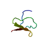
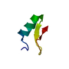
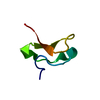
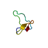
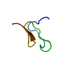
| Title | Exploring Cystine Dense Peptide Space to Open a Unique Molecular Toolbox | ||||||
 Components Components | Chlorotoxin | ||||||
 Keywords Keywords | TOXIN / Knottins / Cystine knot / Toxins | ||||||
| Function / homology | Scorpion short chain toxin, chloride channel inhibitor / Scorpion short toxin / Scorpion short toxin chloride channel inhibitor subfamily profile. / peptidase inhibitor activity / chloride channel regulator activity / Knottin, scorpion toxin-like superfamily / toxin activity / extracellular region / Chlorotoxin Function and homology information Function and homology information | ||||||
| Biological species |  Leiurus quinquestriatus quinquestriatus (Egyptian scorpion) Leiurus quinquestriatus quinquestriatus (Egyptian scorpion) | ||||||
| Method |  X-RAY DIFFRACTION / MOLECULAR REPLACEMENT / Resolution: 1.53 Å X-RAY DIFFRACTION / MOLECULAR REPLACEMENT / Resolution: 1.53 Å | ||||||
 Authors Authors | Gewe, M.M. / Rupert, P. / Strong, R.K. | ||||||
 Citation Citation | Journal: Nat. Struct. Mol. Biol. / Year: 2018 Title: Screening, large-scale production and structure-based classification of cystine-dense peptides. Authors: Correnti, C.E. / Gewe, M.M. / Mehlin, C. / Bandaranayake, A.D. / Johnsen, W.A. / Rupert, P.B. / Brusniak, M.Y. / Clarke, M. / Burke, S.E. / De Van Der Schueren, W. / Pilat, K. / Turnbaugh, S. ...Authors: Correnti, C.E. / Gewe, M.M. / Mehlin, C. / Bandaranayake, A.D. / Johnsen, W.A. / Rupert, P.B. / Brusniak, M.Y. / Clarke, M. / Burke, S.E. / De Van Der Schueren, W. / Pilat, K. / Turnbaugh, S.M. / May, D. / Watson, A. / Chan, M.K. / Bahl, C.D. / Olson, J.M. / Strong, R.K. | ||||||
| History |
|
- Structure visualization
| Structure viewer | Molecule: MolmilJmol/JSmol |
|---|
- Downloads & links
-Download
| PDBx/mmCIF format | 6atw.cif.gz | 20.9 KB | Display | PDBx/mmCIF format |
|---|---|---|---|---|
| PDB format | pdb6atw.ent.gz | 11.4 KB | Display | PDB format |
| PDBx/mmJSON format | 6atw.json.gz | Tree view | PDBx/mmJSON format | |
| Others |  Other downloads Other downloads |
-Validation report
| Arichive directory | https://data.pdbj.org/pub/pdb/validation_reports/at/6atwftp://data.pdbj.org/pub/pdb/validation_reports/at/6atw | HTTPS FTP |
|---|
-Related structure data
| Related structure data |  6atlC  6atnC 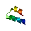 6atsC 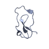 6atuC  6au7C 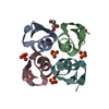 6aupC  6av8C  6avaC  6avcC  6avdC C: citing same article ( |
|---|---|
| Similar structure data |


-Links
 PDBj
PDBj

- Assembly
| Deposited unit | 
| ||||||||
|---|---|---|---|---|---|---|---|---|---|
| 1 |
| ||||||||
| Unit cell |
|
-Components
| #1: Protein/peptide | Mass: 4155.942 Da / Num. of mol.: 1 Source method: isolated from a genetically manipulated source Source: (gene. exp.) Leiurus quinquestriatus quinquestriatus (Egyptian scorpion)Cell (production host): HEK-293F / Production host:  Homo sapiens (human) / References: UniProt: P45639 Homo sapiens (human) / References: UniProt: P45639 |
|---|---|
| #2: Water | ChemComp-HOH /  Mass: 18.015 Da / Num. of mol.: 55 / Source method: isolated from a natural source / Formula: H2O Mass: 18.015 Da / Num. of mol.: 55 / Source method: isolated from a natural source / Formula: H2O |
| Has protein modification | Y |
-Experimental details
-Experiment
| Experiment | Method: X-RAY DIFFRACTION / Number of used crystals: 1 |
|---|
- Sample preparation
| Crystal | Density Matthews: 1.71 Å3/Da / Density % sol: 28.23 % |
|---|---|
| Crystal grow | Temperature: 298 K / Method: vapor diffusion, sitting drop / pH: 7 Details: 1.1 M Na malonate pH 7.0, 0.1 M HEPES pH 7.0, 0.5 % Jeffamine ED-2001 pH 7.0 |
-Data collection
| Diffraction | Mean temperature: 100 K |
|---|---|
| Diffraction source | Source: ROTATING ANODE / Type: RIGAKU MICROMAX-007 HF / Wavelength: 1.54 Å |
| Detector | Type: RIGAKU SATURN 944+ / Detector: CCD / Date: Mar 9, 2016 |
| Radiation | Protocol: SINGLE WAVELENGTH / Monochromatic (M) / Laue (L): M / Scattering type: x-ray |
| Radiation wavelength | Wavelength: 1.54 Å / Relative weight: 1 |
| Reflection | Resolution: 1.53→50 Å / Num. obs: 7263 / % possible obs: 86.9 % / Redundancy: 13.1 % / Rmerge(I) obs: 0.095 / Rpim(I) all: 0.023 / Net I/σ(I): 13.1 |
| Reflection shell | Resolution: 1.53→1.56 Å / Redundancy: 2.3 % / Rmerge(I) obs: 0.126 / Num. unique obs: 46 / CC1/2: 0.935 / Rpim(I) all: 0.076 / % possible all: 10.7 |
- Processing
| Software |
| ||||||||||||||||||||||||||||||||||||||||||||||||||||||||||||||||||||||||||||||||||||||||||||||||||||||||||||||||||||||||||||||||||||||||||||||||||||||||||||||||||||||||||||||||||||||
|---|---|---|---|---|---|---|---|---|---|---|---|---|---|---|---|---|---|---|---|---|---|---|---|---|---|---|---|---|---|---|---|---|---|---|---|---|---|---|---|---|---|---|---|---|---|---|---|---|---|---|---|---|---|---|---|---|---|---|---|---|---|---|---|---|---|---|---|---|---|---|---|---|---|---|---|---|---|---|---|---|---|---|---|---|---|---|---|---|---|---|---|---|---|---|---|---|---|---|---|---|---|---|---|---|---|---|---|---|---|---|---|---|---|---|---|---|---|---|---|---|---|---|---|---|---|---|---|---|---|---|---|---|---|---|---|---|---|---|---|---|---|---|---|---|---|---|---|---|---|---|---|---|---|---|---|---|---|---|---|---|---|---|---|---|---|---|---|---|---|---|---|---|---|---|---|---|---|---|---|---|---|---|---|
| Refinement | Method to determine structure: MOLECULAR REPLACEMENT / Resolution: 1.53→24.02 Å / Cor.coef. Fo:Fc: 0.974 / Cor.coef. Fo:Fc free: 0.948 / SU B: 1.065 / SU ML: 0.04 / Cross valid method: THROUGHOUT / ESU R: 0.081 / ESU R Free: 0.087 / Details: HYDROGENS HAVE BEEN ADDED IN THE RIDING POSITIONS
| ||||||||||||||||||||||||||||||||||||||||||||||||||||||||||||||||||||||||||||||||||||||||||||||||||||||||||||||||||||||||||||||||||||||||||||||||||||||||||||||||||||||||||||||||||||||
| Solvent computation | Ion probe radii: 0.8 Å / Shrinkage radii: 0.8 Å / VDW probe radii: 1.2 Å | ||||||||||||||||||||||||||||||||||||||||||||||||||||||||||||||||||||||||||||||||||||||||||||||||||||||||||||||||||||||||||||||||||||||||||||||||||||||||||||||||||||||||||||||||||||||
| Displacement parameters | Biso mean: 15.516 Å2
| ||||||||||||||||||||||||||||||||||||||||||||||||||||||||||||||||||||||||||||||||||||||||||||||||||||||||||||||||||||||||||||||||||||||||||||||||||||||||||||||||||||||||||||||||||||||
| Refinement step | Cycle: 1 / Resolution: 1.53→24.02 Å
| ||||||||||||||||||||||||||||||||||||||||||||||||||||||||||||||||||||||||||||||||||||||||||||||||||||||||||||||||||||||||||||||||||||||||||||||||||||||||||||||||||||||||||||||||||||||
| Refine LS restraints |
|