 Movie
Movie Controller


+ Open data

- Basic information
| Entry | Database: PDB / ID: 6arh | ||||||
|---|---|---|---|---|---|---|---|
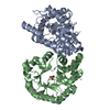
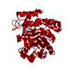
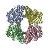
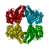
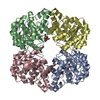
| Title | Crystal structure of Human NAL at a resolution of 1.6 Angstrom | ||||||
 Components Components | N-acetylneuraminate lyase | ||||||
 Keywords Keywords | LYASE / sugar metabolism / sialic acid / tetramer | ||||||
| Function / homology |  Function and homology information Function and homology informationN-acetylneuraminate lyase / N-acetylneuraminate lyase activity / Sialic acid metabolism / N-acetylneuraminate catabolic process / carbohydrate metabolic process / identical protein binding / cytosol Similarity search - Function | ||||||
| Biological species |  Homo sapiens (human) Homo sapiens (human) | ||||||
| Method |  X-RAY DIFFRACTION / SYNCHROTRON / MOLECULAR REPLACEMENT / Resolution: 1.6 Å X-RAY DIFFRACTION / SYNCHROTRON / MOLECULAR REPLACEMENT / Resolution: 1.6 Å | ||||||
 Authors Authors | Pearce, F.G. / Bundela, R. / Keown, J.R. | ||||||
 Citation Citation | Journal: To Be Published Title: Crystal structure of Human NAL at a resolution of 1.6 Angstrom Authors: Pearce, F.G. / Bundela, R. / Keown, J.R. | ||||||
| History |
|
- Structure visualization
| Structure viewer | Molecule: MolmilJmol/JSmol |
|---|
- Downloads & links
-Download
| PDBx/mmCIF format | 6arh.cif.gz | 271.5 KB | Display | PDBx/mmCIF format |
|---|---|---|---|---|
| PDB format | pdb6arh.ent.gz | 215.7 KB | Display | PDB format |
| PDBx/mmJSON format | 6arh.json.gz | Tree view | PDBx/mmJSON format | |
| Others |  Other downloads Other downloads |
-Validation report
| Arichive directory | https://data.pdbj.org/pub/pdb/validation_reports/ar/6arhftp://data.pdbj.org/pub/pdb/validation_reports/ar/6arh | HTTPS FTP |
|---|
-Related structure data
| Related structure data |  5afdS S: Starting model for refinement |
|---|---|
| Similar structure data |


-Links
 PDBj
PDBj- Assembly
| Deposited unit | 
| ||||||||
|---|---|---|---|---|---|---|---|---|---|
| 1 |
| ||||||||
| Unit cell |
|
-Components
| #1: Protein | Mass: 37266.586 Da / Num. of mol.: 4 Source method: isolated from a genetically manipulated source Source: (gene. exp.) Homo sapiens (human) / Gene: NPL, C1orf13 / Production host:  #2: Chemical | ChemComp-ACT /   Mass: 59.044 Da / Num. of mol.: 8 / Source method: obtained synthetically / Formula: C2H3O2 Mass: 59.044 Da / Num. of mol.: 8 / Source method: obtained synthetically / Formula: C2H3O2#3: Water | ChemComp-HOH / |  Mass: 18.015 Da / Num. of mol.: 1271 / Source method: isolated from a natural source / Formula: H2O Mass: 18.015 Da / Num. of mol.: 1271 / Source method: isolated from a natural source / Formula: H2O |
|---|
-Experimental details
-Experiment
| Experiment | Method: X-RAY DIFFRACTION / Number of used crystals: 1 |
|---|
- Sample preparation
| Crystal | Density Matthews: 2.46 Å3/Da / Density % sol: 50.06 % |
|---|---|
| Crystal grow | Temperature: 291 K / Method: vapor diffusion / pH: 7.5 Details: 25% PEG3350, 0.2 M sodium acetate, 0.1 M HEPES, pH 7.5 |
-Data collection
| Diffraction | Mean temperature: 110 K | ||||||||||||||||||||||||
|---|---|---|---|---|---|---|---|---|---|---|---|---|---|---|---|---|---|---|---|---|---|---|---|---|---|
| Diffraction source | Source: SYNCHROTRON / Site: Australian Synchrotron  / Beamline: MX1 / Wavelength: 0.9537 Å / Beamline: MX1 / Wavelength: 0.9537 Å | ||||||||||||||||||||||||
| Detector | Type: ADSC QUANTUM 210r / Detector: CCD / Date: Apr 1, 2017 | ||||||||||||||||||||||||
| Radiation | Monochromator: double crystal Si(111) / Protocol: SINGLE WAVELENGTH / Monochromatic (M) / Laue (L): M / Scattering type: x-ray | ||||||||||||||||||||||||
| Radiation wavelength | Wavelength: 0.9537 Å / Relative weight: 1 | ||||||||||||||||||||||||
| Reflection | Resolution: 1.6→49.27 Å / Num. obs: 173953 / % possible obs: 100 % / Redundancy: 6.8 % / CC1/2: 0.998 / Rmerge(I) obs: 0.09 / Rpim(I) all: 0.037 / Rrim(I) all: 0.097 / Net I/σ(I): 10.4 | ||||||||||||||||||||||||
| Reflection shell | Diffraction-ID: 1
|
- Processing
| Software |
| ||||||||||||||||||||||||||||||||||||||||||||||||||||||||||||
|---|---|---|---|---|---|---|---|---|---|---|---|---|---|---|---|---|---|---|---|---|---|---|---|---|---|---|---|---|---|---|---|---|---|---|---|---|---|---|---|---|---|---|---|---|---|---|---|---|---|---|---|---|---|---|---|---|---|---|---|---|---|
| Refinement | Method to determine structure: MOLECULAR REPLACEMENT Starting model: PDB entry 5AFD Resolution: 1.6→49.27 Å / Cor.coef. Fo:Fc: 0.972 / Cor.coef. Fo:Fc free: 0.963 / SU B: 1.764 / SU ML: 0.059 / SU R Cruickshank DPI: 0.0793 / Cross valid method: THROUGHOUT / σ(F): 0 / ESU R: 0.079 / ESU R Free: 0.078 / Details: HYDROGENS HAVE BEEN ADDED IN THE RIDING POSITIONS
| ||||||||||||||||||||||||||||||||||||||||||||||||||||||||||||
| Solvent computation | Ion probe radii: 0.8 Å / Shrinkage radii: 0.8 Å / VDW probe radii: 1.2 Å | ||||||||||||||||||||||||||||||||||||||||||||||||||||||||||||
| Displacement parameters | Biso max: 68.46 Å2 / Biso mean: 23.337 Å2 / Biso min: 13.99 Å2
| ||||||||||||||||||||||||||||||||||||||||||||||||||||||||||||
| Refinement step | Cycle: final / Resolution: 1.6→49.27 Å
| ||||||||||||||||||||||||||||||||||||||||||||||||||||||||||||
| Refine LS restraints |
| ||||||||||||||||||||||||||||||||||||||||||||||||||||||||||||
| LS refinement shell | Resolution: 1.6→1.642 Å / Rfactor Rfree error: 0 / Total num. of bins used: 20
|
