 Movie
Movie Controller

[English] 日本語
 Yorodumi
- PDB-6aoi: Crystal structure of Toxoplasma gondii TS-DHFR complexed with NAD... -
+ Open data

- Basic information
| Entry | Database: PDB / ID: 6aoi | ||||||
|---|---|---|---|---|---|---|---|
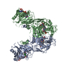
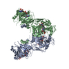
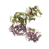
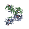
| Title | Crystal structure of Toxoplasma gondii TS-DHFR complexed with NADPH, dUMP, PDDF, and 5-(4-phenylpiperazin-1-yl)-6-propylpyrimidine-2,4-diamine (TRC-2528) | ||||||
 Components Components | Bifunctional dihydrofolate reductase-thymidylate synthase | ||||||
 Keywords Keywords | OXIDOREDUCTASE / Toxoplasma gondii / Pyrimethamine / DHFR | ||||||
| Function / homology |  Function and homology information Function and homology informationthymidylate synthase / thymidylate synthase activity / dTMP biosynthetic process / dihydrofolate reductase / dihydrofolate reductase activity / tetrahydrofolate biosynthetic process / one-carbon metabolic process / methylation / mitochondrion / cytosol Similarity search - Function | ||||||
| Biological species |  | ||||||
| Method |  X-RAY DIFFRACTION / SYNCHROTRON / MOLECULAR REPLACEMENT / Resolution: 2.97 Å X-RAY DIFFRACTION / SYNCHROTRON / MOLECULAR REPLACEMENT / Resolution: 2.97 Å | ||||||
 Authors Authors | Thomas, S.B. / Li, Y. / Chen, Z. / Lu, H. | ||||||
 Citation Citation | Journal: To Be Published Title: Identification of selective, brain penetrant Toxoplasma gondii dihydrofolate reductase inhibitors for the treatment of toxoplasmosis Authors: Thomas, S.B. / Hopper, A. | ||||||
| History |
|
- Structure visualization
| Structure viewer | Molecule: MolmilJmol/JSmol |
|---|
- Downloads & links
-Download
| PDBx/mmCIF format | 6aoi.cif.gz | 225.2 KB | Display | PDBx/mmCIF format |
|---|---|---|---|---|
| PDB format | pdb6aoi.ent.gz | 178.2 KB | Display | PDB format |
| PDBx/mmJSON format | 6aoi.json.gz | Tree view | PDBx/mmJSON format | |
| Others |  Other downloads Other downloads |
-Validation report
| Arichive directory | https://data.pdbj.org/pub/pdb/validation_reports/ao/6aoiftp://data.pdbj.org/pub/pdb/validation_reports/ao/6aoi | HTTPS FTP |
|---|
-Related structure data
| Related structure data |  6aogC 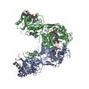 6aohC  4eilS C: citing same article ( S: Starting model for refinement |
|---|---|
| Similar structure data |


-Links
 PDBj
PDBj

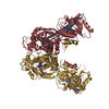
- Assembly
| Deposited unit | 
| ||||||||
|---|---|---|---|---|---|---|---|---|---|
| 1 |
| ||||||||
| Unit cell |
|
-Components
| #1: Protein | Mass: 64214.312 Da / Num. of mol.: 2 Source method: isolated from a genetically manipulated source Source: (gene. exp.)  References: UniProt: Q07422, dihydrofolate reductase, thymidylate synthase #2: Chemical |   Mass: 308.182 Da / Num. of mol.: 2 Mass: 308.182 Da / Num. of mol.: 2Source method: isolated from a genetically manipulated source Formula: C9H13N2O8P #3: Chemical |   Mass: 745.421 Da / Num. of mol.: 2 Mass: 745.421 Da / Num. of mol.: 2Source method: isolated from a genetically manipulated source Formula: C21H30N7O17P3 #4: Chemical |   Mass: 477.469 Da / Num. of mol.: 2 Mass: 477.469 Da / Num. of mol.: 2Source method: isolated from a genetically manipulated source Formula: C24H23N5O6 #5: Chemical |   Mass: 312.413 Da / Num. of mol.: 2 / Source method: obtained synthetically / Formula: C17H24N6 Mass: 312.413 Da / Num. of mol.: 2 / Source method: obtained synthetically / Formula: C17H24N6 |
|---|
-Experimental details
-Experiment
| Experiment | Method: X-RAY DIFFRACTION / Number of used crystals: 1 |
|---|
- Sample preparation
| Crystal | Density Matthews: 2.47 Å3/Da / Density % sol: 50.13 % |
|---|---|
| Crystal grow | Temperature: 293 K / Method: vapor diffusion, sitting drop / pH: 4.8 / Details: 0.2 M ammonium citrate, pH 4.6, 15% PEG3350 |
-Data collection
| Diffraction | Mean temperature: 100 K |
|---|---|
| Diffraction source | Source: SYNCHROTRON / Site: SSRF  / Beamline: BL17U1 / Wavelength: 0.979282 Å / Beamline: BL17U1 / Wavelength: 0.979282 Å |
| Detector | Type: ADSC QUANTUM 315r / Detector: CCD / Date: Sep 26, 2016 |
| Radiation | Protocol: SINGLE WAVELENGTH / Monochromatic (M) / Laue (L): M / Scattering type: x-ray |
| Radiation wavelength | Wavelength: 0.979282 Å / Relative weight: 1 |
| Reflection | Resolution: 2.97→50 Å / Num. obs: 26892 / % possible obs: 91.7 % / Redundancy: 2.7 % / Rmerge(I) obs: 0.114 / Net I/σ(I): 9.8 |
| Reflection shell | Resolution: 2.97→3.08 Å / Rmerge(I) obs: 0.596 |
- Processing
| Software |
| ||||||||||||||||||||||||||||||||||||||||||||||||||||||||||||||||||||||||||||||||||||||||||||||||||||||||||||||||||||||||||||||||||||||||||||||||||||||||||||||||||||||||||||||||||||||
|---|---|---|---|---|---|---|---|---|---|---|---|---|---|---|---|---|---|---|---|---|---|---|---|---|---|---|---|---|---|---|---|---|---|---|---|---|---|---|---|---|---|---|---|---|---|---|---|---|---|---|---|---|---|---|---|---|---|---|---|---|---|---|---|---|---|---|---|---|---|---|---|---|---|---|---|---|---|---|---|---|---|---|---|---|---|---|---|---|---|---|---|---|---|---|---|---|---|---|---|---|---|---|---|---|---|---|---|---|---|---|---|---|---|---|---|---|---|---|---|---|---|---|---|---|---|---|---|---|---|---|---|---|---|---|---|---|---|---|---|---|---|---|---|---|---|---|---|---|---|---|---|---|---|---|---|---|---|---|---|---|---|---|---|---|---|---|---|---|---|---|---|---|---|---|---|---|---|---|---|---|---|---|---|
| Refinement | Method to determine structure: MOLECULAR REPLACEMENT Starting model: PDB entry 4EIL Resolution: 2.97→50 Å / Cor.coef. Fo:Fc: 0.932 / Cor.coef. Fo:Fc free: 0.886 / Cross valid method: THROUGHOUT / ESU R Free: 0.49
| ||||||||||||||||||||||||||||||||||||||||||||||||||||||||||||||||||||||||||||||||||||||||||||||||||||||||||||||||||||||||||||||||||||||||||||||||||||||||||||||||||||||||||||||||||||||
| Solvent computation | Ion probe radii: 0.8 Å / Shrinkage radii: 0.8 Å / VDW probe radii: 1.2 Å | ||||||||||||||||||||||||||||||||||||||||||||||||||||||||||||||||||||||||||||||||||||||||||||||||||||||||||||||||||||||||||||||||||||||||||||||||||||||||||||||||||||||||||||||||||||||
| Displacement parameters | Biso mean: 83.287 Å2
| ||||||||||||||||||||||||||||||||||||||||||||||||||||||||||||||||||||||||||||||||||||||||||||||||||||||||||||||||||||||||||||||||||||||||||||||||||||||||||||||||||||||||||||||||||||||
| Refinement step | Cycle: 1 / Resolution: 2.97→50 Å
| ||||||||||||||||||||||||||||||||||||||||||||||||||||||||||||||||||||||||||||||||||||||||||||||||||||||||||||||||||||||||||||||||||||||||||||||||||||||||||||||||||||||||||||||||||||||
| Refine LS restraints |
|