 Movie
Movie Controller

[English] 日本語
 Yorodumi
- PDB-6alw: The crystal structure of the Staphylococcus aureus Fatty acid Kin... -
+ Open data

- Basic information
| Entry | Database: PDB / ID: 6alw | ||||||
|---|---|---|---|---|---|---|---|
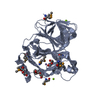
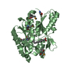
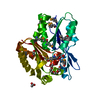
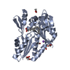
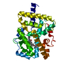
| Title | The crystal structure of the Staphylococcus aureus Fatty acid Kinase (Fak) B1 protein loaded with 12-Methyl Myristic Acid (C15:0) to 1.63 Angstrom resolution | ||||||
 Components Components | EDD domain protein, DegV family | ||||||
 Keywords Keywords | TRANSFERASE / Staphylococcus aureus / FakB1 / 12-Methyl Myristic Acid / 12-Methyltetradecanoic acid / C15:0 | ||||||
| Function / homology |  Function and homology information Function and homology information | ||||||
| Biological species |   Staphylococcus aureus (bacteria) Staphylococcus aureus (bacteria) | ||||||
| Method |  X-RAY DIFFRACTION / SYNCHROTRON / MOLECULAR REPLACEMENT / Resolution: 1.63 Å X-RAY DIFFRACTION / SYNCHROTRON / MOLECULAR REPLACEMENT / Resolution: 1.63 Å | ||||||
 Authors Authors | Cuypers, M.G. / Ericson, M. / Subramanian, C. / White, S.W. / Rock, C.O. | ||||||
| Funding support |  United States, 1items United States, 1items
| ||||||
 Citation Citation | Journal: J. Biol. Chem. / Year: 2019 Title: Acyl-chain selectivity and physiological roles ofStaphylococcus aureusfatty acid-binding proteins. Authors: Cuypers, M.G. / Subramanian, C. / Gullett, J.M. / Frank, M.W. / White, S.W. / Rock, C.O. | ||||||
| History |
|
- Structure visualization
| Structure viewer | Molecule: MolmilJmol/JSmol |
|---|
- Downloads & links
-Download
| PDBx/mmCIF format | 6alw.cif.gz | 279.5 KB | Display | PDBx/mmCIF format |
|---|---|---|---|---|
| PDB format | pdb6alw.ent.gz | 225.9 KB | Display | PDB format |
| PDBx/mmJSON format | 6alw.json.gz | Tree view | PDBx/mmJSON format | |
| Others |  Other downloads Other downloads |
-Validation report
| Arichive directory | https://data.pdbj.org/pub/pdb/validation_reports/al/6alwftp://data.pdbj.org/pub/pdb/validation_reports/al/6alw | HTTPS FTP |
|---|
-Related structure data
| Related structure data |  5wooC 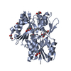 6b9iC 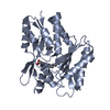 5utoS C: citing same article ( S: Starting model for refinement |
|---|---|
| Similar structure data |


-Links
 PDBj
PDBj- Assembly
| Deposited unit | 
| ||||||||
|---|---|---|---|---|---|---|---|---|---|
| 1 | 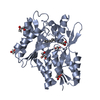
| ||||||||
| 2 | 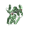
| ||||||||
| Unit cell |
|
-Components
| #1: Protein | Mass: 32101.506 Da / Num. of mol.: 2 Source method: isolated from a genetically manipulated source Source: (gene. exp.) Staphylococcus aureus (bacteria)Gene: AYM28_04055, AYM37_04055, ERS072738_00223, ERS072840_01626, ERS074020_00218, HMPREF3211_01094 Production host: #2: Chemical |   Mass: 242.397 Da / Num. of mol.: 2 / Source method: obtained synthetically / Formula: C15H30O2 Mass: 242.397 Da / Num. of mol.: 2 / Source method: obtained synthetically / Formula: C15H30O2#3: Chemical |   Mass: 242.397 Da / Num. of mol.: 2 / Source method: obtained synthetically / Formula: C15H30O2 Mass: 242.397 Da / Num. of mol.: 2 / Source method: obtained synthetically / Formula: C15H30O2#4: Chemical | ChemComp-GOL /   Mass: 92.094 Da / Num. of mol.: 8 / Source method: obtained synthetically / Formula: C3H8O3 Mass: 92.094 Da / Num. of mol.: 8 / Source method: obtained synthetically / Formula: C3H8O3#5: Water | ChemComp-HOH / |  Mass: 18.015 Da / Num. of mol.: 838 / Source method: isolated from a natural source / Formula: H2O Mass: 18.015 Da / Num. of mol.: 838 / Source method: isolated from a natural source / Formula: H2O |
|---|
-Experimental details
-Experiment
| Experiment | Method: X-RAY DIFFRACTION / Number of used crystals: 1 |
|---|
- Sample preparation
| Crystal | Density Matthews: 2.19 Å3/Da / Density % sol: 43.81 % |
|---|---|
| Crystal grow | Temperature: 293 K / Method: vapor diffusion, hanging drop Details: PH 6.5 0.1M MES/IMIDAZOLE, 12.5% PEG1000, 12.5% PEG3350, 12.5% MPD, 0.03M NaNO3, 0.03M NA2HPO4, 0.03M (NH4)2 SO4 Temp details: controlled room temperature |
-Data collection
| Diffraction | Mean temperature: 100 K / Ambient temp details: crystal flash frozen in Liq. N2 |
|---|---|
| Diffraction source | Source: SYNCHROTRON / Site: APS / Beamline: 22-BM / Wavelength: 1 Å |
| Detector | Type: MARMOSAIC 225 mm CCD / Detector: CCD / Date: Nov 10, 2016 |
| Radiation | Protocol: SINGLE WAVELENGTH / Monochromatic (M) / Laue (L): M / Scattering type: x-ray |
| Radiation wavelength | Wavelength: 1 Å / Relative weight: 1 |
| Reflection | Resolution: 1.63→81.18 Å / Num. obs: 66112 / % possible obs: 97.2 % / Redundancy: 4 % / Biso Wilson estimate: 7.76 Å2 / CC1/2: 0.985 / Rmerge(I) obs: 0.184 / Rpim(I) all: 0.106 / Rrim(I) all: 0.212 / Net I/σ(I): 8.6 |
| Reflection shell | Resolution: 1.63→1.66 Å / Redundancy: 4 % / Rmerge(I) obs: 0.906 / Mean I/σ(I) obs: 2 / Num. unique obs: 3294 / CC1/2: 0.662 / Rpim(I) all: 0.524 / % possible all: 95.2 |
- Processing
| Software |
| |||||||||||||||||||||||||||||||||||||||||||||||||||||||||||||||||||||||||||||||||||||||||||||||||||||||||||||||||||||||||||||||||||||||||||||||||||||||||||||||||||||||||||||||
|---|---|---|---|---|---|---|---|---|---|---|---|---|---|---|---|---|---|---|---|---|---|---|---|---|---|---|---|---|---|---|---|---|---|---|---|---|---|---|---|---|---|---|---|---|---|---|---|---|---|---|---|---|---|---|---|---|---|---|---|---|---|---|---|---|---|---|---|---|---|---|---|---|---|---|---|---|---|---|---|---|---|---|---|---|---|---|---|---|---|---|---|---|---|---|---|---|---|---|---|---|---|---|---|---|---|---|---|---|---|---|---|---|---|---|---|---|---|---|---|---|---|---|---|---|---|---|---|---|---|---|---|---|---|---|---|---|---|---|---|---|---|---|---|---|---|---|---|---|---|---|---|---|---|---|---|---|---|---|---|---|---|---|---|---|---|---|---|---|---|---|---|---|---|---|---|---|
| Refinement | Method to determine structure: MOLECULAR REPLACEMENT Starting model: 5UTO Resolution: 1.63→40.591 Å / SU ML: 0.18 / Cross valid method: FREE R-VALUE / σ(F): 1.98 / Phase error: 19.42 / Stereochemistry target values: ML
| |||||||||||||||||||||||||||||||||||||||||||||||||||||||||||||||||||||||||||||||||||||||||||||||||||||||||||||||||||||||||||||||||||||||||||||||||||||||||||||||||||||||||||||||
| Solvent computation | Shrinkage radii: 0.9 Å / VDW probe radii: 1.11 Å / Solvent model: FLAT BULK SOLVENT MODEL | |||||||||||||||||||||||||||||||||||||||||||||||||||||||||||||||||||||||||||||||||||||||||||||||||||||||||||||||||||||||||||||||||||||||||||||||||||||||||||||||||||||||||||||||
| Refinement step | Cycle: LAST / Resolution: 1.63→40.591 Å
| |||||||||||||||||||||||||||||||||||||||||||||||||||||||||||||||||||||||||||||||||||||||||||||||||||||||||||||||||||||||||||||||||||||||||||||||||||||||||||||||||||||||||||||||
| Refine LS restraints |
| |||||||||||||||||||||||||||||||||||||||||||||||||||||||||||||||||||||||||||||||||||||||||||||||||||||||||||||||||||||||||||||||||||||||||||||||||||||||||||||||||||||||||||||||
| LS refinement shell |
| |||||||||||||||||||||||||||||||||||||||||||||||||||||||||||||||||||||||||||||||||||||||||||||||||||||||||||||||||||||||||||||||||||||||||||||||||||||||||||||||||||||||||||||||
| Refinement TLS params. | Method: refined / Origin x: 0.2649 Å / Origin y: -0.9655 Å / Origin z: -0.289 Å
| |||||||||||||||||||||||||||||||||||||||||||||||||||||||||||||||||||||||||||||||||||||||||||||||||||||||||||||||||||||||||||||||||||||||||||||||||||||||||||||||||||||||||||||||
| Refinement TLS group | Selection details: all |
