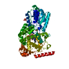
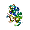
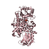
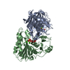
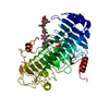
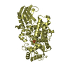
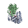
Entry Database : PDB / ID : 6akdTitle Crystal structure of IdnL7 AMP-dependent synthetase and ligase Keywords / / Function / homology Function Domain/homology Component
/ / / / / / / / / Biological species Streptomyces sp. ML694-90F3 (bacteria)Method / / / Resolution : 2.1 Å Authors Cieslak, J. / Miyanaga, A. / Kudo, F. / Eguchi, T. Funding support Organization Grant number Country Japan Society for the Promotion of Science 25850050
Journal : Acta Crystallogr F Struct Biol Commun / Year : 2019Title : Functional and structural characterization of IdnL7, an adenylation enzyme involved in incednine biosynthesis.Authors : Cieslak, J. / Miyanaga, A. / Takaishi, M. / Kudo, F. / Eguchi, T. History Deposition Aug 31, 2018 Deposition site / Processing site Revision 1.0 Mar 6, 2019 Provider / Type Revision 1.1 Apr 17, 2019 Group / Database references / Category / citation_authorItem _citation.journal_abbrev / _citation.journal_volume ... _citation.journal_abbrev / _citation.journal_volume / _citation.page_first / _citation.page_last / _citation.pdbx_database_id_DOI / _citation.pdbx_database_id_PubMed / _citation.title / _citation_author.identifier_ORCID Revision 1.2 Nov 22, 2023 Group / Database references / Refinement descriptionCategory chem_comp_atom / chem_comp_bond ... chem_comp_atom / chem_comp_bond / database_2 / pdbx_initial_refinement_model Item / _database_2.pdbx_database_accession
Show all Show less
 Movie
Movie Controller


 Open data
 Basic information
 Components
 Keywords
 Function and homology information
 Streptomyces sp. ML694-90F3 (bacteria)
 X-RAY DIFFRACTION /
  Authors
 Japan, 1items
  Citation
 Structure visualization
 Downloads & links
 Other downloads
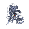


 PDBj
PDBj
 Assembly
 Mass: 417.398 Da / Num. of mol.: 1 / Source method: obtained synthetically / Formula: C13H19N7O7S
 Mass: 92.094 Da / Num. of mol.: 2 / Source method: obtained synthetically / Formula: C3H8O3
 Mass: 18.015 Da / Num. of mol.: 155 / Source method: isolated from a natural source / Formula: H2O
 Sample preparation
 Processing