[English] 日本語
Yorodumi
- PDB-3ief: Crystal structure of tRNA guanine-n1-methyltransferase from Barto... -

+
Open data


ID or keywords:

Loading...

-
Basic information

Entry
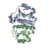
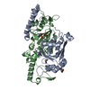
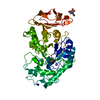
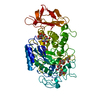
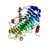
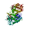
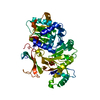
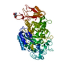
Database: PDB / ID: 3ief
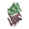
TitleCrystal structure of tRNA guanine-n1-methyltransferase from Bartonella henselae using MPCS.
ComponentstRNA (guanine-N(1)-)-methyltransferase
KeywordsTRANSFERASE / RNA BINDING PROTEIN / NIAID / SSGCID / Seattle Structural Genomics Center for Infectious Diseases / Microcapillary Protein Crystallization System / MPCS / PSI-2 / Protein Structure Initiative / Accelerated Technologies Center for Gene to 3D Structure / ATCG3D / Cytoplasm / Methyltransferase / S-adenosyl-L-methionine / tRNA processing / protein knot
Function / homology
Function and homology information


tRNA (guanine37-N1)-methyltransferase / tRNA (guanine(37)-N1)-methyltransferase activity / tRNA modification / methylation / cytoplasm
Similarity search - Function
tRNA(m1g37)methyltransferase, domain 2 / Trp Operon Repressor; Chain A / tRNA (guanine-N1-)-methyltransferase, bacteria / tRNA (guanine-N(1)-)-methyltransferase, C-terminal domain superfamily / tRNA methyltransferase TRMD/TRM10-type domain / tRNA (Guanine-1)-methyltransferase / SPOUT methyltransferase, trefoil knot domain / Alpha/beta knot / tRNA (guanine-N1-)-methyltransferase, N-terminal / Alpha/beta knot methyltransferases ...tRNA(m1g37)methyltransferase, domain 2 / Trp Operon Repressor; Chain A / tRNA (guanine-N1-)-methyltransferase, bacteria / tRNA (guanine-N(1)-)-methyltransferase, C-terminal domain superfamily / tRNA methyltransferase TRMD/TRM10-type domain / tRNA (Guanine-1)-methyltransferase / SPOUT methyltransferase, trefoil knot domain / Alpha/beta knot / tRNA (guanine-N1-)-methyltransferase, N-terminal / Alpha/beta knot methyltransferases / Orthogonal Bundle / 3-Layer(aba) Sandwich / Mainly Alpha / Alpha Beta
Similarity search - Domain/homology
tRNA (guanine-N(1)-)-methyltransferase
Similarity search - Component
Biological speciesBartonella henselae (bacteria)
MethodX-RAY DIFFRACTION / SYNCHROTRON / MOLECULAR REPLACEMENT / molecular replacement / Resolution: 2.5 Å
AuthorsEdwards, T.E. / Stahl, G. / Gerdts, C.J. / Staker, B.L. / Accelerated Technologies Center for Gene to 3D Structure (ATCG3D) / Seattle Structural Genomics Center for Infectious Disease (SSGCID)
CitationJournal: To be Published
Title: Crystal structure of tRNA guanine-n1-methyltransferase from Bartonella henselae using MPCS.
Authors: Edwards, T.E. / Stahl, G. / Gerdts, C.J. / Staker, B.L. / Accelerated Technologies Center for Gene to 3D Structure (ATCG3D) / Seattle Structural Genomics Center for Infectious Disease (SSGCID)
History
DepositionJul 22, 2009Deposition site: RCSB / Processing site: RCSB
Revision 1.0Aug 11, 2009Provider: repository / Type: Initial release
Revision 1.1Jul 13, 2011Group: Version format compliance
Revision 1.2Nov 1, 2017Group: Refinement description / Category: software
Revision 1.3Sep 6, 2023Group: Data collection / Database references / Refinement description
Category: chem_comp_atom / chem_comp_bond ...chem_comp_atom / chem_comp_bond / database_2 / pdbx_initial_refinement_model
Item: _database_2.pdbx_DOI / _database_2.pdbx_database_accession

-
Structure visualization

Structure viewerMolecule:
MolmilJmol/JSmol

Downloads & links

-
Assembly

Deposited unit
A: tRNA (guanine-N(1)-)-methyltransferase
B: tRNA (guanine-N(1)-)-methyltransferase


Theoretical massNumber of molelcules
Total (without water)51,1622
Polymers51,1622
Non-polymers00
Water1,67593
1


  • Idetical with deposited unit
  • defined by software
TypeNameSymmetry operationNumber
identity operation1_555x,y,z1
Buried area6650 Å2
ΔGint-21 kcal/mol
Surface area19170 Å2
MethodPISA
Unit cell
Length a, b, c (Å)56.017, 84.467, 97.192
Angle α, β, γ (deg.)90.000, 90.000, 90.000
Int Tables number19
Space group name H-MP212121

-
Components

#1: Protein tRNA (guanine-N(1)-)-methyltransferase / M1G-methyltransferase / tRNA [GM37] methyltransferase


Mass: 25581.215 Da / Num. of mol.: 2
Source method: isolated from a genetically manipulated source
Details: Expressed as a fusion protein with H-terminal hexahis tag and Smt3 protein. Cleaved with ULP-1 protease, then purified as target protein with only one Ser from expression tag.
Source: (gene. exp.) Bartonella henselae (bacteria) / Strain: Houston-1 / Gene: trmD, BH15820 / Plasmid: pBADSmt / Production host: Escherichia coli (E. coli) / References: UniProt: Q6G1R9, EC: 2.1.1.31
#2: Water ChemComp-HOH / water


Mass: 18.015 Da / Num. of mol.: 93 / Source method: isolated from a natural source / Formula: H2O

-
Experimental details

-
Experiment

ExperimentMethod: X-RAY DIFFRACTION / Number of used crystals: 1

-
Sample preparation

CrystalDensity Matthews: 2.25 Å3/Da / Density % sol: 45.26 %
Crystal growTemperature: 293 K / Method: mpcs / pH: 7
Details: Initial crystal hit JCSG+ condition G7 15% PEG 3350, 0.1M succinic acid pH 7.0 optimized with microcapillary gradient, MPCS, temperature 293K

-
Data collection

DiffractionMean temperature: 100 K
Diffraction sourceSource: SYNCHROTRON / Site: SSRL / Beamline: BL7-1 / Wavelength: 0.97946 Å
DetectorDate: Jun 19, 2009
RadiationProtocol: SINGLE WAVELENGTH / Monochromatic (M) / Laue (L): M / Scattering type: x-ray
Radiation wavelengthWavelength: 0.97946 Å / Relative weight: 1
ReflectionResolution: 2.5→50 Å / Num. obs: 16569 / % possible obs: 99.9 % / Redundancy: 8.9 % / Rmerge(I) obs: 0.116 / Χ2: 0.997 / Net I/σ(I): 18.2
Reflection shellResolution: 2.5→2.59 Å / Redundancy: 8.5 % / Rmerge(I) obs: 0.492 / Mean I/σ(I) obs: 3.8 / Num. unique all: 1614 / Χ2: 0.987 / % possible all: 100

-
Phasing

PhasingMethod: molecular replacement
Phasing MRRfactor: 50.97 / Model details: Phaser MODE: MR_AUTO
Highest resolutionLowest resolution
Rotation3 Å38.73 Å
Translation3 Å38.73 Å

-
Processing

Software
NameVersionClassificationNB
DENZOdata reduction
SCALEPACKdata scaling
PHASER2.1.4phasing
REFMACrefinement
PDB_EXTRACT3.005data extraction
HKL-2000data reduction
HKL-2000data scaling
RefinementMethod to determine structure: MOLECULAR REPLACEMENT
Starting model: 1p9p
Resolution: 2.5→50 Å / Cor.coef. Fo:Fc: 0.938 / Cor.coef. Fo:Fc free: 0.936 / WRfactor Rfree: 0.199 / WRfactor Rwork: 0.173 / Occupancy max: 1 / Occupancy min: 1 / FOM work R set: 0.847 / SU B: 8.475 / SU ML: 0.191 / SU R Cruickshank DPI: 0.619 / SU Rfree: 0.266 / Cross valid method: THROUGHOUT / σ(F): 0 / ESU R: 0.619 / ESU R Free: 0.266 / Stereochemistry target values: MAXIMUM LIKELIHOOD
Details: HYDROGENS HAVE BEEN ADDED IN THE RIDING POSITIONS U VALUES : REFINED INDIVIDUALLY
RfactorNum. reflection% reflectionSelection details
Rfree0.225 825 5 %RANDOM
Rwork0.195 ---
obs0.196 16525 99.89 %-
Solvent computationIon probe radii: 0.8 Å / Shrinkage radii: 0.8 Å / VDW probe radii: 1.4 Å / Solvent model: MASK
Displacement parametersBiso max: 67.43 Å2 / Biso mean: 30.164 Å2 / Biso min: 8.67 Å2
Baniso -1Baniso -2Baniso -3
1--0.99 Å20 Å20 Å2
2---1.98 Å20 Å2
3---2.97 Å2
Refinement stepCycle: LAST / Resolution: 2.5→50 Å
ProteinNucleic acidLigandSolventTotal
Num. atoms3287 0 0 93 3380
Refine LS restraints
Refine-IDTypeDev idealDev ideal targetNumber
X-RAY DIFFRACTIONr_bond_refined_d0.0130.0223349
X-RAY DIFFRACTIONr_angle_refined_deg1.3811.9784544
X-RAY DIFFRACTIONr_dihedral_angle_1_deg6.0395434
X-RAY DIFFRACTIONr_dihedral_angle_2_deg33.11222.897145
X-RAY DIFFRACTIONr_dihedral_angle_3_deg15.45515526
X-RAY DIFFRACTIONr_dihedral_angle_4_deg18.3051534
X-RAY DIFFRACTIONr_chiral_restr0.0840.2514
X-RAY DIFFRACTIONr_gen_planes_refined0.0060.0212569
X-RAY DIFFRACTIONr_mcbond_it0.781.52173
X-RAY DIFFRACTIONr_mcangle_it1.47123445
X-RAY DIFFRACTIONr_scbond_it1.99931176
X-RAY DIFFRACTIONr_scangle_it3.2964.51099
LS refinement shellResolution: 2.5→2.564 Å / Total num. of bins used: 20
RfactorNum. reflection% reflection
Rfree0.324 49 -
Rwork0.239 1136 -
all-1185 -
obs--99.33 %

+
About Yorodumi

-
News

-
Feb 9, 2022. New format data for meta-information of EMDB entries

New format data for meta-information of EMDB entries

  • Version 3 of the EMDB header file is now the official format.
  • The previous official version 1.9 will be removed from the archive.

Related info.:EMDB header

External links:wwPDB to switch to version 3 of the EMDB data model

-
Aug 12, 2020. Covid-19 info

Covid-19 info

URL: https://pdbj.org/emnavi/covid19.php

New page: Covid-19 featured information page in EM Navigator.

Related info.:Covid-19 info / Mar 5, 2020. Novel coronavirus structure data

+
Mar 5, 2020. Novel coronavirus structure data

Novel coronavirus structure data

Related info.:Yorodumi Speices / Aug 12, 2020. Covid-19 info

External links:COVID-19 featured content - PDBj / Molecule of the Month (242):Coronavirus Proteases

+
Jan 31, 2019. EMDB accession codes are about to change! (news from PDBe EMDB page)

EMDB accession codes are about to change! (news from PDBe EMDB page)

  • The allocation of 4 digits for EMDB accession codes will soon come to an end. Whilst these codes will remain in use, new EMDB accession codes will include an additional digit and will expand incrementally as the available range of codes is exhausted. The current 4-digit format prefixed with “EMD-” (i.e. EMD-XXXX) will advance to a 5-digit format (i.e. EMD-XXXXX), and so on. It is currently estimated that the 4-digit codes will be depleted around Spring 2019, at which point the 5-digit format will come into force.
  • The EM Navigator/Yorodumi systems omit the EMD- prefix.

Related info.:Q: What is EMD? / ID/Accession-code notation in Yorodumi/EM Navigator

External links:EMDB Accession Codes are Changing Soon! / Contact to PDBj

+
Jul 12, 2017. Major update of PDB

Major update of PDB

  • wwPDB released updated PDB data conforming to the new PDBx/mmCIF dictionary.
  • This is a major update changing the version number from 4 to 5, and with Remediation, in which all the entries are updated.
  • In this update, many items about electron microscopy experimental information are reorganized (e.g. em_software).
  • Now, EM Navigator and Yorodumi are based on the updated data.

External links:wwPDB Remediation / Enriched Model Files Conforming to OneDep Data Standards Now Available in the PDB FTP Archive

-
Yorodumi

Thousand views of thousand structures

  • Yorodumi is a browser for structure data from EMDB, PDB, SASBDB, etc.
  • This page is also the successor to EM Navigator detail page, and also detail information page/front-end page for Omokage search.
  • The word "yorodu" (or yorozu) is an old Japanese word meaning "ten thousand". "mi" (miru) is to see.

Related info.:EMDB / PDB / SASBDB / Comparison of 3 databanks / Yorodumi Search / Aug 31, 2016. New EM Navigator & Yorodumi / Yorodumi Papers / Jmol/JSmol / Function and homology information / Changes in new EM Navigator and Yorodumi

Read more