 Movie
Movie Controller

[English] 日本語
 Yorodumi
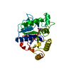
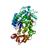
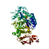
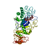
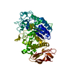
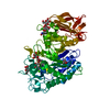
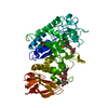
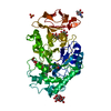
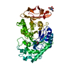
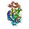
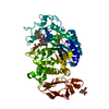
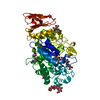
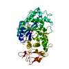
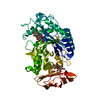
- PDB-1kgw: THREE DIMENSIONAL STRUCTURE ANALYSIS OF THE R337Q VARIANT OF HUMA... -
+ Open data

- Basic information
| Entry | Database: PDB / ID: 1kgw | |||||||||
|---|---|---|---|---|---|---|---|---|---|---|
| Title | THREE DIMENSIONAL STRUCTURE ANALYSIS OF THE R337Q VARIANT OF HUMAN PANCREATIC ALPHA-MYLASE | |||||||||
 Components Components | ALPHA-AMYLASE, PANCREATIC | |||||||||
 Keywords Keywords | HYDROLASE / ALPHA-AMYLASE / CHLORIDE BINDING / PICHIA PASTORIS / MUTAGENESIS / CATALYSIS / PANCREATIC / ENZYME / HUMAN | |||||||||
| Function / homology |  Function and homology information Function and homology informationpolysaccharide digestion / Digestion of dietary carbohydrate / Developmental Lineage of Pancreatic Acinar Cells / alpha-amylase / alpha-amylase activity / carbohydrate catabolic process / chloride ion binding / carbohydrate metabolic process / calcium ion binding / : ...polysaccharide digestion / Digestion of dietary carbohydrate / Developmental Lineage of Pancreatic Acinar Cells / alpha-amylase / alpha-amylase activity / carbohydrate catabolic process / chloride ion binding / carbohydrate metabolic process / calcium ion binding / : / extracellular exosome / extracellular region Similarity search - Function | |||||||||
| Biological species |  Homo sapiens (human) Homo sapiens (human) | |||||||||
| Method |  X-RAY DIFFRACTION / MOLECULAR REPLACEMENT / Resolution: 2.1 Å X-RAY DIFFRACTION / MOLECULAR REPLACEMENT / Resolution: 2.1 Å | |||||||||
 Authors Authors | Numao, S. / Maurus, R. / Sidhu, G. / Wang, Y. / Overall, C.M. / Brayer, G.D. / Withers, S.G. | |||||||||
 Citation Citation | Journal: Biochemistry / Year: 2002 Title: Probing the role of the chloride ion in the mechanism of human pancreatic alpha-amylase. Authors: Numao, S. / Maurus, R. / Sidhu, G. / Wang, Y. / Overall, C.M. / Brayer, G.D. / Withers, S.G. | |||||||||
| History |
|
- Structure visualization
| Structure viewer | Molecule: MolmilJmol/JSmol |
|---|
- Downloads & links
-Download
| PDBx/mmCIF format | 1kgw.cif.gz | 114.7 KB | Display | PDBx/mmCIF format |
|---|---|---|---|---|
| PDB format | pdb1kgw.ent.gz | 86.7 KB | Display | PDB format |
| PDBx/mmJSON format | 1kgw.json.gz | Tree view | PDBx/mmJSON format | |
| Others |  Other downloads Other downloads |
-Validation report
| Arichive directory | https://data.pdbj.org/pub/pdb/validation_reports/kg/1kgwftp://data.pdbj.org/pub/pdb/validation_reports/kg/1kgw | HTTPS FTP |
|---|
-Related structure data
| Related structure data |  1kb3C 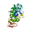 1kguC 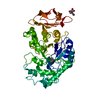 1kgxC  1bsiS C: citing same article ( S: Starting model for refinement |
|---|---|
| Similar structure data |


-Links
 PDBj
PDBj

- Assembly
| Deposited unit | 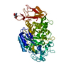
| ||||||||
|---|---|---|---|---|---|---|---|---|---|
| 1 |
| ||||||||
| Unit cell |
|
-Components
| #1: Protein | Mass: 55902.238 Da / Num. of mol.: 1 / Mutation: R377Q Source method: isolated from a genetically manipulated source Source: (gene. exp.) Homo sapiens (human) / Production host:  Pichia pastoris (fungus) / References: UniProt: P04746, alpha-amylase Pichia pastoris (fungus) / References: UniProt: P04746, alpha-amylase |
|---|---|
| #2: Sugar | ChemComp-NAG /   Type: D-saccharide, beta linking / Mass: 221.208 Da / Num. of mol.: 1 Type: D-saccharide, beta linking / Mass: 221.208 Da / Num. of mol.: 1Source method: isolated from a genetically manipulated source Formula: C8H15NO6 |
| #3: Chemical | ChemComp-CA /   Mass: 40.078 Da / Num. of mol.: 1 / Source method: obtained synthetically / Formula: Ca Mass: 40.078 Da / Num. of mol.: 1 / Source method: obtained synthetically / Formula: Ca |
| #4: Water | ChemComp-HOH /  Mass: 18.015 Da / Num. of mol.: 171 / Source method: isolated from a natural source / Formula: H2O Mass: 18.015 Da / Num. of mol.: 171 / Source method: isolated from a natural source / Formula: H2O |
| Has protein modification | Y |
-Experimental details
-Experiment
| Experiment | Method: X-RAY DIFFRACTION / Number of used crystals: 1 |
|---|
- Sample preparation
| Crystal | Density Matthews: 2.43 Å3/Da / Density % sol: 49.39 % | ||||||||||||||||||||
|---|---|---|---|---|---|---|---|---|---|---|---|---|---|---|---|---|---|---|---|---|---|
| Crystal grow | Temperature: 298 K / Method: vapor diffusion, hanging drop / Details: VAPOR DIFFUSION, HANGING DROP, temperature 298K | ||||||||||||||||||||
| Crystal grow | *PLUS pH: 7.5 / Details: Brayer, G.D., (2000) Biochemistry, 39, 4778. | ||||||||||||||||||||
| Components of the solutions | *PLUS
|
-Data collection
| Diffraction | Mean temperature: 298 K |
|---|---|
| Diffraction source | Source: ROTATING ANODE / Type: RIGAKU RU300 / Wavelength: 1.5418 / Wavelength: 1.5418 Å |
| Detector | Type: RIGAKU RAXIS IIC / Detector: IMAGE PLATE / Date: Jul 15, 1999 / Details: MIRRORS |
| Radiation | Monochromator: MIRRORS / Protocol: SINGLE WAVELENGTH / Monochromatic (M) / Laue (L): M / Scattering type: x-ray |
| Radiation wavelength | Wavelength: 1.5418 Å / Relative weight: 1 |
| Reflection | Resolution: 2→30 Å / Num. all: 29654 / Num. obs: 29654 / Observed criterion σ(I): 0 / Redundancy: 3.8 % / Rmerge(I) obs: 0.056 / Net I/σ(I): 17.3 |
| Reflection | *PLUS Highest resolution: 2 Å / Lowest resolution: 30 Å / Redundancy: 3.8 % / Num. measured all: 297678 |
| Reflection shell | *PLUS Highest resolution: 2.1 Å / Lowest resolution: 2.14 Å / Redundancy: 2.2 % / Rmerge(I) obs: 0.134 / Mean I/σ(I) obs: 8.2 |
- Processing
| Software |
| ||||||||||||||||||
|---|---|---|---|---|---|---|---|---|---|---|---|---|---|---|---|---|---|---|---|
| Refinement | Method to determine structure: MOLECULAR REPLACEMENT Starting model: PDB ENTRY 1BSI Resolution: 2.1→10 Å / Isotropic thermal model: ISOTROPIC / σ(F): 0 / σ(I): 0 / Stereochemistry target values: Engh & Huber
| ||||||||||||||||||
| Displacement parameters | Biso mean: 22.6 Å2 | ||||||||||||||||||
| Refine analyze | Luzzati coordinate error obs: 0.23 Å | ||||||||||||||||||
| Refinement step | Cycle: LAST / Resolution: 2.1→10 Å
| ||||||||||||||||||
| Software | *PLUS Name: X-PLOR / Version: 3.851 / Classification: refinement | ||||||||||||||||||
| Refinement | *PLUS Rfactor obs: 0.19 | ||||||||||||||||||
| Solvent computation | *PLUS | ||||||||||||||||||
| Displacement parameters | *PLUS | ||||||||||||||||||
| Refine LS restraints | *PLUS
|
