 Movie
Movie Controller

[English] 日本語
 Yorodumi
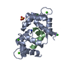
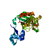
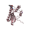
- PDB-6aeq: Crystal structure of the ssDNA-binding domain of DnaT from Salmon... -
+ Open data

- Basic information
| Entry | Database: PDB / ID: 6aeq | ||||||
|---|---|---|---|---|---|---|---|
| Title | Crystal structure of the ssDNA-binding domain of DnaT from Salmonella enterica Serovar Typhimurium LT2 | ||||||
 Components Components | Primosomal protein 1 | ||||||
 Keywords Keywords | DNA BINDING PROTEIN / Primosome / replication restart / DnaT / DNA binding | ||||||
| Function / homology |  Function and homology information Function and homology informationprimosome complex / DNA replication, synthesis of primer / single-stranded DNA binding Similarity search - Function | ||||||
| Biological species |  Salmonella typhimurium (bacteria) Salmonella typhimurium (bacteria) | ||||||
| Method |  X-RAY DIFFRACTION / SYNCHROTRON / MOLECULAR REPLACEMENT / Resolution: 2.2518084706 Å X-RAY DIFFRACTION / SYNCHROTRON / MOLECULAR REPLACEMENT / Resolution: 2.2518084706 Å | ||||||
 Authors Authors | Huang, Y.H. / Huang, C.Y. | ||||||
 Citation Citation | Journal: Biochem. Biophys. Res. Commun. / Year: 2019 Title: Crystal structure of the C-terminal domain of the primosomal DnaT protein: Insights into a new oligomerization mechanism. Authors: Chen, K.L. / Huang, Y.H. / Liao, J.F. / Lee, W.C. / Huang, C.Y. | ||||||
| History |
|
- Structure visualization
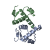
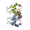
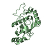
| Structure viewer | Molecule: MolmilJmol/JSmol |
|---|
- Downloads & links
-Download
| PDBx/mmCIF format | 6aeq.cif.gz | 52.7 KB | Display | PDBx/mmCIF format |
|---|---|---|---|---|
| PDB format | pdb6aeq.ent.gz | 30.4 KB | Display | PDB format |
| PDBx/mmJSON format | 6aeq.json.gz | Tree view | PDBx/mmJSON format | |
| Others |  Other downloads Other downloads |
-Validation report
| Arichive directory | https://data.pdbj.org/pub/pdb/validation_reports/ae/6aeqftp://data.pdbj.org/pub/pdb/validation_reports/ae/6aeq | HTTPS FTP |
|---|
-Related structure data
| Related structure data | 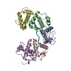 4ou6S S: Starting model for refinement |
|---|---|
| Similar structure data |


-Links
 PDBj
PDBj- Assembly
| Deposited unit | 
| |||||||||||||||||||||
|---|---|---|---|---|---|---|---|---|---|---|---|---|---|---|---|---|---|---|---|---|---|---|
| 1 |
| |||||||||||||||||||||
| Unit cell |
| |||||||||||||||||||||
| Noncrystallographic symmetry (NCS) | NCS domain:
NCS domain segments: Ens-ID: 1 / Beg auth comp-ID: ILE / Beg label comp-ID: ILE / End auth comp-ID: ASN / End label comp-ID: ASN / Auth seq-ID: 84 - 155 / Label seq-ID: 1 - 72
|
-Components
| #1: Protein | Mass: 11646.032 Da / Num. of mol.: 2 Source method: isolated from a genetically manipulated source Source: (gene. exp.) Salmonella typhimurium (strain LT2 / SGSC1412 / ATCC 700720) (bacteria)Strain: LT2 / SGSC1412 / ATCC 700720 / Gene: dnaT, STM4544 / Production host: #2: Water | ChemComp-HOH / |  Mass: 18.015 Da / Num. of mol.: 16 / Source method: isolated from a natural source / Formula: H2O Mass: 18.015 Da / Num. of mol.: 16 / Source method: isolated from a natural source / Formula: H2OHas protein modification | Y | |
|---|
-Experimental details
-Experiment
| Experiment | Method: X-RAY DIFFRACTION / Number of used crystals: 1 |
|---|
- Sample preparation
| Crystal | Density Matthews: 1.88 Å3/Da / Density % sol: 34.48 % |
|---|---|
| Crystal grow | Temperature: 298 K / Method: vapor diffusion, hanging drop / pH: 6.5 Details: 1.6M Magnesium Sulfate, 100mM MES Sodium Salt pH 6.5 |
-Data collection
| Diffraction | Mean temperature: 298 K |
|---|---|
| Diffraction source | Source: SYNCHROTRON / Site: NSRRC  / Beamline: BL13B1 / Wavelength: 1 Å / Beamline: BL13B1 / Wavelength: 1 Å |
| Detector | Type: ADSC QUANTUM 315 / Detector: CCD / Date: May 10, 2018 |
| Radiation | Protocol: SINGLE WAVELENGTH / Monochromatic (M) / Laue (L): M / Scattering type: x-ray |
| Radiation wavelength | Wavelength: 1 Å / Relative weight: 1 |
| Reflection | Resolution: 2.25→30 Å / Num. obs: 8294 / % possible obs: 98.4 % / Redundancy: 5.6 % / Rmerge(I) obs: 0.052 / Net I/σ(I): 30.14 |
| Reflection shell | Resolution: 2.25→2.38 Å / Redundancy: 5 % / Rmerge(I) obs: 0.288 / Mean I/σ(I) obs: 4.58 / Num. unique obs: 764 / % possible all: 91.8 |
- Processing
| Software |
| ||||||||||||||||||||||||||||
|---|---|---|---|---|---|---|---|---|---|---|---|---|---|---|---|---|---|---|---|---|---|---|---|---|---|---|---|---|---|
| Refinement | Method to determine structure: MOLECULAR REPLACEMENT Starting model: 4OU6 Resolution: 2.2518084706→21.48309024 Å / SU ML: 0.235716035848 / Cross valid method: THROUGHOUT / σ(F): 1.33784763378 / Phase error: 34.1707603168
| ||||||||||||||||||||||||||||
| Solvent computation | Shrinkage radii: 0.9 Å / VDW probe radii: 1.11 Å | ||||||||||||||||||||||||||||
| Displacement parameters | Biso mean: 54.3122439788 Å2 | ||||||||||||||||||||||||||||
| Refinement step | Cycle: LAST / Resolution: 2.2518084706→21.48309024 Å
| ||||||||||||||||||||||||||||
| Refine LS restraints |
| ||||||||||||||||||||||||||||
| LS refinement shell |
|
