 Movie
Movie Controller

[English] 日本語
 Yorodumi
- PDB-6aa8: Crystal structure of (S)-3-hydroxybutyryl-coenzymeA dehydrogenase... -
+ Open data

- Basic information
| Entry | Database: PDB / ID: 6aa8 | ||||||
|---|---|---|---|---|---|---|---|
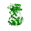
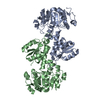
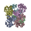
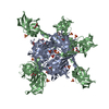
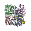
| Title | Crystal structure of (S)-3-hydroxybutyryl-coenzymeA dehydrogenase from Clostridium acetobutylicum complexed with NAD+ | ||||||
 Components Components | 3-hydroxybutyryl-CoA dehydrogenase | ||||||
 Keywords Keywords | OXIDOREDUCTASE / Complex / Rossmann fold | ||||||
| Function / homology |  Function and homology information Function and homology information3-hydroxybutyryl-CoA dehydrogenase / butyrate metabolic process / 3-hydroxybutyryl-CoA dehydrogenase activity / fatty acid beta-oxidation / NAD+ binding Similarity search - Function | ||||||
| Biological species |  Clostridium acetobutylicum (bacteria) Clostridium acetobutylicum (bacteria) | ||||||
| Method |  X-RAY DIFFRACTION / SYNCHROTRON / MOLECULAR REPLACEMENT / Resolution: 2.1 Å X-RAY DIFFRACTION / SYNCHROTRON / MOLECULAR REPLACEMENT / Resolution: 2.1 Å | ||||||
 Authors Authors | Takenoya, M. / Taguchi, S. / Yajima, S. | ||||||
 Citation Citation | Journal: Acta Crystallogr F Struct Biol Commun / Year: 2018 Title: Crystal structure and kinetic analyses of a hexameric form of (S)-3-hydroxybutyryl-CoA dehydrogenase from Clostridium acetobutylicum. Authors: Takenoya, M. / Taguchi, S. / Yajima, S. | ||||||
| History |
|
- Structure visualization
| Structure viewer | Molecule: MolmilJmol/JSmol |
|---|
- Downloads & links
-Download
| PDBx/mmCIF format | 6aa8.cif.gz | 328.6 KB | Display | PDBx/mmCIF format |
|---|---|---|---|---|
| PDB format | pdb6aa8.ent.gz | 265.9 KB | Display | PDB format |
| PDBx/mmJSON format | 6aa8.json.gz | Tree view | PDBx/mmJSON format | |
| Others |  Other downloads Other downloads |
-Validation report
| Arichive directory | https://data.pdbj.org/pub/pdb/validation_reports/aa/6aa8ftp://data.pdbj.org/pub/pdb/validation_reports/aa/6aa8 | HTTPS FTP |
|---|
-Related structure data
| Related structure data |  6acqC 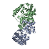 4r1nS S: Starting model for refinement C: citing same article ( |
|---|---|
| Similar structure data |

-Links
 PDBj
PDBj


- Assembly
| Deposited unit | 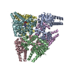
| ||||||||
|---|---|---|---|---|---|---|---|---|---|
| 1 |
| ||||||||
| Unit cell |
|
-Components
| #1: Protein | Mass: 32793.043 Da / Num. of mol.: 6 Source method: isolated from a genetically manipulated source Source: (gene. exp.) Clostridium acetobutylicum (strain ATCC 824 / DSM 792 / JCM 1419 / LMG 5710 / VKM B-1787) (bacteria)Strain: ATCC 824 / DSM 792 / JCM 1419 / LMG 5710 / VKM B-1787 Gene: hbd, CA_C2708 / Production host: References: UniProt: P52041, 3-hydroxybutyryl-CoA dehydrogenase #2: Chemical |   Mass: 663.425 Da / Num. of mol.: 2 / Source method: obtained synthetically / Formula: C21H27N7O14P2 / Comment: NAD*YM Mass: 663.425 Da / Num. of mol.: 2 / Source method: obtained synthetically / Formula: C21H27N7O14P2 / Comment: NAD*YM#3: Water | ChemComp-HOH / |  Mass: 18.015 Da / Num. of mol.: 404 / Source method: isolated from a natural source / Formula: H2O Mass: 18.015 Da / Num. of mol.: 404 / Source method: isolated from a natural source / Formula: H2O |
|---|
-Experimental details
-Experiment
| Experiment | Method: X-RAY DIFFRACTION / Number of used crystals: 1 |
|---|
- Sample preparation
| Crystal | Density Matthews: 2.69 Å3/Da / Density % sol: 54.32 % |
|---|---|
| Crystal grow | Temperature: 293 K / Method: vapor diffusion, hanging drop / Details: 17.5% PEG 3350, 200mM sodium thiocyanate |
-Data collection
| Diffraction | Mean temperature: 100 K |
|---|---|
| Diffraction source | Source: SYNCHROTRON / Site: Photon Factory  / Beamline: BL-17A / Wavelength: 0.98 Å / Beamline: BL-17A / Wavelength: 0.98 Å |
| Detector | Type: ADSC QUANTUM 270 / Detector: CCD / Date: Feb 26, 2018 |
| Radiation | Protocol: SINGLE WAVELENGTH / Monochromatic (M) / Laue (L): M / Scattering type: x-ray |
| Radiation wavelength | Wavelength: 0.98 Å / Relative weight: 1 |
| Reflection | Resolution: 2.1→50 Å / Num. obs: 109647 / % possible obs: 98.6 % / Redundancy: 2.2 % / CC1/2: 0.996 / Rmerge(I) obs: 0.049 / Net I/σ(I): 14.1 |
| Reflection shell | Resolution: 2.1→2.14 Å / Rmerge(I) obs: 0.316 / Num. unique obs: 5492 / CC1/2: 0.829 |
- Processing
| Software |
| ||||||||||||||||||||||||||||||||||||||||||||||||||||||||||||||||||||||||||||||||||||||||||||||||||||||||||||||||||||||||||||||||||||||||||||||||||||||||||||||||||||||||||||||||||||||
|---|---|---|---|---|---|---|---|---|---|---|---|---|---|---|---|---|---|---|---|---|---|---|---|---|---|---|---|---|---|---|---|---|---|---|---|---|---|---|---|---|---|---|---|---|---|---|---|---|---|---|---|---|---|---|---|---|---|---|---|---|---|---|---|---|---|---|---|---|---|---|---|---|---|---|---|---|---|---|---|---|---|---|---|---|---|---|---|---|---|---|---|---|---|---|---|---|---|---|---|---|---|---|---|---|---|---|---|---|---|---|---|---|---|---|---|---|---|---|---|---|---|---|---|---|---|---|---|---|---|---|---|---|---|---|---|---|---|---|---|---|---|---|---|---|---|---|---|---|---|---|---|---|---|---|---|---|---|---|---|---|---|---|---|---|---|---|---|---|---|---|---|---|---|---|---|---|---|---|---|---|---|---|---|
| Refinement | Method to determine structure: MOLECULAR REPLACEMENT Starting model: 4R1N Resolution: 2.1→48.46 Å / Cor.coef. Fo:Fc: 0.955 / Cor.coef. Fo:Fc free: 0.94 / SU B: 4.701 / SU ML: 0.123 / Cross valid method: THROUGHOUT / ESU R: 0.211 / ESU R Free: 0.172 / Stereochemistry target values: MAXIMUM LIKELIHOOD / Details: HYDROGENS HAVE BEEN ADDED IN THE RIDING POSITIONS
| ||||||||||||||||||||||||||||||||||||||||||||||||||||||||||||||||||||||||||||||||||||||||||||||||||||||||||||||||||||||||||||||||||||||||||||||||||||||||||||||||||||||||||||||||||||||
| Solvent computation | Ion probe radii: 0.8 Å / Shrinkage radii: 0.8 Å / VDW probe radii: 1.2 Å / Solvent model: MASK | ||||||||||||||||||||||||||||||||||||||||||||||||||||||||||||||||||||||||||||||||||||||||||||||||||||||||||||||||||||||||||||||||||||||||||||||||||||||||||||||||||||||||||||||||||||||
| Displacement parameters | Biso mean: 36.829 Å2
| ||||||||||||||||||||||||||||||||||||||||||||||||||||||||||||||||||||||||||||||||||||||||||||||||||||||||||||||||||||||||||||||||||||||||||||||||||||||||||||||||||||||||||||||||||||||
| Refinement step | Cycle: 1 / Resolution: 2.1→48.46 Å
| ||||||||||||||||||||||||||||||||||||||||||||||||||||||||||||||||||||||||||||||||||||||||||||||||||||||||||||||||||||||||||||||||||||||||||||||||||||||||||||||||||||||||||||||||||||||
| Refine LS restraints |
|