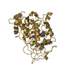
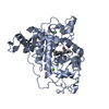
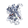
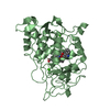
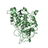
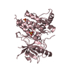
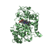
登録情報 データベース : PDB / ID : 6a6qタイトル Crystal structure of a lignin peroxidase isozyme H8 variant that is stable at very acidic pH Ligninase H8 キーワード / / 機能・相同性 分子機能 ドメイン・相同性 構成要素
/ / / / / / / / / / / / / / / / / / / / / / / / / / / / / / / / / 生物種 Phanerochaete chrysosporium RP-78 (菌類)手法 / / / 解像度 : 1.67 Å データ登録者 Seo, H. / Kim, K.-J. / Pham, L.T.M. ジャーナル : Biotechnol Biofuels / 年 : 2018タイトル : In silico-designed lignin peroxidase fromPhanerochaete chrysosporiumshows enhanced acid stability for depolymerization of lignin.著者 : Pham, L.T.M. / Seo, H. / Kim, K.J. / Kim, Y.H. 履歴 登録 2018年6月29日 登録サイト / 処理サイト 改定 1.0 2019年1月23日 Provider / タイプ 改定 1.1 2020年9月16日 Group / Structure summaryカテゴリ pdbx_struct_conn_angle / struct ... pdbx_struct_conn_angle / struct / struct_conn / struct_conn_type Item _pdbx_struct_conn_angle.ptnr1_auth_seq_id / _pdbx_struct_conn_angle.ptnr3_auth_seq_id ... _pdbx_struct_conn_angle.ptnr1_auth_seq_id / _pdbx_struct_conn_angle.ptnr3_auth_seq_id / _pdbx_struct_conn_angle.value / _struct.title / _struct_conn.conn_type_id / _struct_conn.id / _struct_conn.pdbx_dist_value / _struct_conn.pdbx_leaving_atom_flag / _struct_conn.ptnr1_auth_comp_id / _struct_conn.ptnr1_auth_seq_id / _struct_conn.ptnr1_label_atom_id / _struct_conn.ptnr1_label_comp_id / _struct_conn.ptnr1_label_seq_id / _struct_conn.ptnr2_auth_comp_id / _struct_conn.ptnr2_auth_seq_id / _struct_conn.ptnr2_label_asym_id / _struct_conn.ptnr2_label_atom_id / _struct_conn.ptnr2_label_comp_id / _struct_conn.ptnr2_label_seq_id / _struct_conn_type.id 改定 1.2 2023年11月22日 Group / Database references / Refinement descriptionカテゴリ chem_comp_atom / chem_comp_bond ... chem_comp_atom / chem_comp_bond / database_2 / pdbx_initial_refinement_model / pdbx_validate_chiral Item / _database_2.pdbx_database_accession
すべて表示 表示を減らす
 ムービー
ムービー コントローラー


 データを開く
 基本情報
基本情報 要素
要素 キーワード
キーワード 機能・相同性情報
 Phanerochaete chrysosporium RP-78 (菌類)
 X線回折 /
  データ登録者
 引用
引用 構造の表示
構造の表示 ダウンロードとリンク
 その他のダウンロード
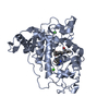


 PDBj
PDBj

 集合体
集合体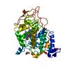


 分子量: 618.503 Da / 分子数: 1 / 由来タイプ: 合成 / 式: C34H34FeN4O4
 分子量: 40.078 Da / 分子数: 2 / 由来タイプ: 合成 / 式: Ca
 分子量: 92.094 Da / 分子数: 3 / 由来タイプ: 合成 / 式: C3H8O3
 分子量: 18.015 Da / 分子数: 249 / 由来タイプ: 天然 / 式: H2O
 試料調製
試料調製 / ビームライン: 7A (6B, 6C1) / 波長: 0.9793 Å
 解析
解析