 Movie
Movie Controller


+ Open data

- Basic information
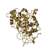
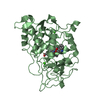
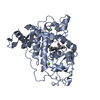
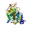
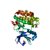
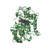
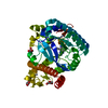
| Entry | Database: PDB / ID: 1b82 | ||||||
|---|---|---|---|---|---|---|---|
| Title | PRISTINE RECOMB. LIGNIN PEROXIDASE H8 | ||||||
 Components Components | PROTEIN (LIGNIN PEROXIDASE) | ||||||
 Keywords Keywords | OXIDOREDUCTASE / LIGNIN DEGRADATION / HEME / RADICAL REACTION / ELECTRON TRANSFER | ||||||
| Function / homology |  Function and homology information Function and homology informationlignin peroxidase / diarylpropane peroxidase activity / lignin catabolic process / response to reactive oxygen species / hydrogen peroxide catabolic process / cellular response to oxidative stress / heme binding / metal ion binding Similarity search - Function | ||||||
| Biological species |  Phanerochaete chrysosporium (fungus) Phanerochaete chrysosporium (fungus) | ||||||
| Method |  X-RAY DIFFRACTION / SYNCHROTRON / MOLECULAR REPLACEMENT / Resolution: 1.8 Å X-RAY DIFFRACTION / SYNCHROTRON / MOLECULAR REPLACEMENT / Resolution: 1.8 Å | ||||||
 Authors Authors | Blodig, W. / Doyle, W.A. / Smith, A.T. / Piontek, K. | ||||||
 Citation Citation | Journal: J.Mol.Biol. / Year: 2001 Title: Crystal structures of pristine and oxidatively processed lignin peroxidase expressed in Escherichia coli and of the W171F variant that eliminates the redox active tryptophan 171. Implications ...Title: Crystal structures of pristine and oxidatively processed lignin peroxidase expressed in Escherichia coli and of the W171F variant that eliminates the redox active tryptophan 171. Implications for the reaction mechanism. Authors: Blodig, W. / Smith, A.T. / Doyle, W.A. / Piontek, K. #1: Journal: Biochemistry / Year: 1998 Title: Two substrate interaction sites in lignin peroxidase revealed by site-directed mutagenesis. Authors: Doyle, W.A. / Blodig, W. / Veitch, N.C. / Piontek, K. / Smith, A.T. #2: Journal: Biochemistry / Year: 1998 Title: Autocatalytic formation of a hydroxy group at C beta of trp171 in lignin peroxidase. Authors: Blodig, W. / Doyle, W.A. / Smith, A.T. / Winterhalter, K. / Choinowski, T. / Piontek, K. #3: Journal: Biochem.J. / Year: 1996 Title: Expression of lignin peroxidase H8 in Escherichia coli: folding and activation of the recombinant enzyme with Ca2+ and haem. Authors: Doyle, W.A. / Smith, A.T. | ||||||
| History |
|
- Structure visualization
| Structure viewer | Molecule: MolmilJmol/JSmol |
|---|
- Downloads & links
-Download
| PDBx/mmCIF format | 1b82.cif.gz | 158.1 KB | Display | PDBx/mmCIF format |
|---|---|---|---|---|
| PDB format | pdb1b82.ent.gz | 122.7 KB | Display | PDB format |
| PDBx/mmJSON format | 1b82.json.gz | Tree view | PDBx/mmJSON format | |
| Others |  Other downloads Other downloads |
-Validation report
| Arichive directory | https://data.pdbj.org/pub/pdb/validation_reports/b8/1b82ftp://data.pdbj.org/pub/pdb/validation_reports/b8/1b82 | HTTPS FTP |
|---|
-Related structure data
| Related structure data | 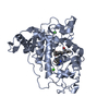 1b80SC  1b85C S: Starting model for refinement C: citing same article ( |
|---|---|
| Similar structure data |


-Links
 PDBj
PDBj
- Assembly
| Deposited unit | 
| ||||||||||
|---|---|---|---|---|---|---|---|---|---|---|---|
| 1 | 
| ||||||||||
| 2 | 
| ||||||||||
| Unit cell |
| ||||||||||
| Noncrystallographic symmetry (NCS) | NCS oper: (Code: given Matrix: (0.258, -0.966, -0.008), Vector: |
-Components
| #1: Protein | Mass: 37480.719 Da / Num. of mol.: 2 / Fragment: MATURE PROTEIN PLUS 7-RESIDUE PROSEQUENCE Source method: isolated from a genetically manipulated source Details: HEME CONTAINING / Source: (gene. exp.) Phanerochaete chrysosporium (fungus) / Strain: BKM 1767 / Description: RECOMBINANT EXPRESSION IN E. COLI / Gene: LIP H8 / Variant: WILD TYPE / Plasmid: PFLAG1-LIPP / Production host:  #2: Chemical | ChemComp-CA /   Mass: 40.078 Da / Num. of mol.: 4 / Source method: obtained synthetically / Formula: Ca Mass: 40.078 Da / Num. of mol.: 4 / Source method: obtained synthetically / Formula: Ca#3: Chemical |   Mass: 616.487 Da / Num. of mol.: 2 / Source method: obtained synthetically / Formula: C34H32FeN4O4 Mass: 616.487 Da / Num. of mol.: 2 / Source method: obtained synthetically / Formula: C34H32FeN4O4#4: Water | ChemComp-HOH / |  Mass: 18.015 Da / Num. of mol.: 548 / Source method: isolated from a natural source / Formula: H2O Mass: 18.015 Da / Num. of mol.: 548 / Source method: isolated from a natural source / Formula: H2OHas protein modification | Y | |
|---|
-Experimental details
-Experiment
| Experiment | Method: X-RAY DIFFRACTION / Number of used crystals: 1 |
|---|
- Sample preparation
| Crystal | Density Matthews: 2.6 Å3/Da / Density % sol: 53 % | |||||||||||||||||||||||||
|---|---|---|---|---|---|---|---|---|---|---|---|---|---|---|---|---|---|---|---|---|---|---|---|---|---|---|
| Crystal grow | pH: 3.5 / Details: pH 3.5 | |||||||||||||||||||||||||
| Components of the solutions |
| |||||||||||||||||||||||||
| Crystal grow | *PLUS Temperature: 20 ℃ / pH: 6 / Method: vapor diffusion, hanging drop | |||||||||||||||||||||||||
| Components of the solutions | *PLUS
|
-Data collection
| Diffraction | Mean temperature: 293 K |
|---|---|
| Diffraction source | Source: SYNCHROTRON / Site: EMBL/DESY, HAMBURG  / Beamline: BW7B / Wavelength: 0.8345 / Beamline: BW7B / Wavelength: 0.8345 |
| Detector | Type: MARRESEARCH / Detector: IMAGE PLATE / Date: Aug 15, 1998 |
| Radiation | Protocol: SINGLE WAVELENGTH / Monochromatic (M) / Laue (L): M / Scattering type: x-ray |
| Radiation wavelength | Wavelength: 0.8345 Å / Relative weight: 1 |
| Reflection | Resolution: 1.8→30 Å / Num. obs: 69484 / % possible obs: 94.7 % / Redundancy: 3.4 % / Biso Wilson estimate: 18.7 Å2 / Rmerge(I) obs: 0.042 / Net I/σ(I): 20 |
| Reflection shell | Resolution: 1.8→1.83 Å / Rmerge(I) obs: 0.195 / Mean I/σ(I) obs: 4.3 / % possible all: 89.9 |
| Reflection | *PLUS Lowest resolution: 30 Å / Num. measured all: 234766 |
| Reflection shell | *PLUS % possible obs: 89.9 % |
- Processing
| Software |
| ||||||||||||||||||||||||||||||||||||||||||||||||||||||||||||||||||||||||||||||||||||
|---|---|---|---|---|---|---|---|---|---|---|---|---|---|---|---|---|---|---|---|---|---|---|---|---|---|---|---|---|---|---|---|---|---|---|---|---|---|---|---|---|---|---|---|---|---|---|---|---|---|---|---|---|---|---|---|---|---|---|---|---|---|---|---|---|---|---|---|---|---|---|---|---|---|---|---|---|---|---|---|---|---|---|---|---|---|
| Refinement | Method to determine structure: MOLECULAR REPLACEMENT Starting model: 1B80 Resolution: 1.8→30 Å / SU B: 2.16 / Cross valid method: THROUGHOUT / σ(F): 0 Details: ANISOTROPIC SCALING (REFMAC) WAS USED TO ACCOUNT FOR CRYSTAL ANISOTROPICITY PRISTINE LIGNIN PEROXIDASE HAS NOT REACTED WITH PEROXIDES. CRYSTALLISATION AND DATA COLLECTION WERE DONE IN THE ...Details: ANISOTROPIC SCALING (REFMAC) WAS USED TO ACCOUNT FOR CRYSTAL ANISOTROPICITY PRISTINE LIGNIN PEROXIDASE HAS NOT REACTED WITH PEROXIDES. CRYSTALLISATION AND DATA COLLECTION WERE DONE IN THE PRESENCE OF THE PHENOLIC ANTIOXIDANT ORCINOL.
| ||||||||||||||||||||||||||||||||||||||||||||||||||||||||||||||||||||||||||||||||||||
| Displacement parameters | Biso mean: 25.6 Å2 | ||||||||||||||||||||||||||||||||||||||||||||||||||||||||||||||||||||||||||||||||||||
| Refinement step | Cycle: LAST / Resolution: 1.8→30 Å
| ||||||||||||||||||||||||||||||||||||||||||||||||||||||||||||||||||||||||||||||||||||
| Refine LS restraints |
| ||||||||||||||||||||||||||||||||||||||||||||||||||||||||||||||||||||||||||||||||||||
| Software | *PLUS Name: REFMAC / Classification: refinement | ||||||||||||||||||||||||||||||||||||||||||||||||||||||||||||||||||||||||||||||||||||
| Refinement | *PLUS Highest resolution: 1.8 Å / Lowest resolution: 30 Å / σ(F): 0 / % reflection Rfree: 5 % / Rfactor obs: 0.163 | ||||||||||||||||||||||||||||||||||||||||||||||||||||||||||||||||||||||||||||||||||||
| Solvent computation | *PLUS | ||||||||||||||||||||||||||||||||||||||||||||||||||||||||||||||||||||||||||||||||||||
| Displacement parameters | *PLUS Biso mean: 25.6 Å2 | ||||||||||||||||||||||||||||||||||||||||||||||||||||||||||||||||||||||||||||||||||||
| Refine LS restraints | *PLUS
| ||||||||||||||||||||||||||||||||||||||||||||||||||||||||||||||||||||||||||||||||||||
| LS refinement shell | *PLUS Rfactor Rfree: 0.254 / Rfactor obs: 0.189 |
