 Movie
Movie Controller

[English] 日本語
 Yorodumi
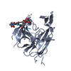
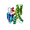
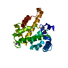
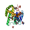
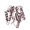
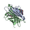
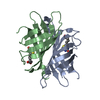
- PDB-5zyv: Crystal structure of human MGME1 with single strand DNA2 and Ca2+ -
+ Open data

- Basic information
| Entry | Database: PDB / ID: 5zyv | ||||||
|---|---|---|---|---|---|---|---|
| Title | Crystal structure of human MGME1 with single strand DNA2 and Ca2+ | ||||||
 Components Components |
| ||||||
 Keywords Keywords | DNA BINDING PROTEIN/DNA / humanMGME1 / DNA complex / DNA nuclease / DNA BINDING PROTEIN / DNA BINDING PROTEIN-DNA complex | ||||||
| Function / homology |  Function and homology information Function and homology informationsingle-stranded DNA exodeoxyribonuclease activity / single-stranded DNA 5'-3' DNA exonuclease activity / Strand-asynchronous mitochondrial DNA replication / mitochondrial DNA replication / 5'-flap endonuclease activity / mitochondrial DNA repair / Hydrolases; Acting on ester bonds / mitochondrial matrix / mitochondrion Similarity search - Function | ||||||
| Biological species |  Homo sapiens (human) Homo sapiens (human)unidentified (others) | ||||||
| Method |  X-RAY DIFFRACTION / SYNCHROTRON / MOLECULAR REPLACEMENT / Resolution: 2.72 Å X-RAY DIFFRACTION / SYNCHROTRON / MOLECULAR REPLACEMENT / Resolution: 2.72 Å | ||||||
 Authors Authors | Yang, C. / Gan, J. | ||||||
 Citation Citation | Journal: Nucleic Acids Res. / Year: 2018 Title: Structural insights into DNA degradation by human mitochondrial nuclease MGME1 Authors: Yang, C. / Wu, R. / Liu, H. / Chen, Y. / Gao, Y. / Chen, X. / Li, Y. / Ma, J. / Li, J. / Gan, J. | ||||||
| History |
|
- Structure visualization
| Structure viewer | Molecule: MolmilJmol/JSmol |
|---|
- Downloads & links
-Download
| PDBx/mmCIF format | 5zyv.cif.gz | 105.5 KB | Display | PDBx/mmCIF format |
|---|---|---|---|---|
| PDB format | pdb5zyv.ent.gz | 76.5 KB | Display | PDB format |
| PDBx/mmJSON format | 5zyv.json.gz | Tree view | PDBx/mmJSON format | |
| Others |  Other downloads Other downloads |
-Validation report
| Arichive directory | https://data.pdbj.org/pub/pdb/validation_reports/zy/5zyvftp://data.pdbj.org/pub/pdb/validation_reports/zy/5zyv | HTTPS FTP |
|---|
-Related structure data
| Related structure data |  5zytSC 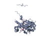 5zyuC 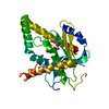 5zywC S: Starting model for refinement C: citing same article ( |
|---|---|
| Similar structure data |


-Links
 PDBj
PDBj


- Assembly
| Deposited unit | 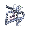
| ||||||||
|---|---|---|---|---|---|---|---|---|---|
| 1 |
| ||||||||
| Unit cell |
|
-Components
| #1: Protein | Mass: 29507.502 Da / Num. of mol.: 1 / Mutation: H180Q Source method: isolated from a genetically manipulated source Source: (gene. exp.) Homo sapiens (human) / Gene: MGME1, C20orf72, DDK1 / Production host:  References: UniProt: Q9BQP7, Hydrolases; Acting on ester bonds | ||||
|---|---|---|---|---|---|
| #2: DNA chain | Mass: 4533.020 Da / Num. of mol.: 1 / Source method: obtained synthetically / Source: (synth.) unidentified (others) | ||||
| #3: Chemical |   Mass: 40.078 Da / Num. of mol.: 2 / Source method: obtained synthetically / Formula: Ca Mass: 40.078 Da / Num. of mol.: 2 / Source method: obtained synthetically / Formula: Ca#4: Chemical | ChemComp-ACT / |   Mass: 59.044 Da / Num. of mol.: 1 / Source method: obtained synthetically / Formula: C2H3O2 Mass: 59.044 Da / Num. of mol.: 1 / Source method: obtained synthetically / Formula: C2H3O2#5: Water | ChemComp-HOH / |  Mass: 18.015 Da / Num. of mol.: 13 / Source method: isolated from a natural source / Formula: H2O Mass: 18.015 Da / Num. of mol.: 13 / Source method: isolated from a natural source / Formula: H2O |
-Experimental details
-Experiment
| Experiment | Method: X-RAY DIFFRACTION / Number of used crystals: 1 |
|---|
- Sample preparation
| Crystal | Density Matthews: 2.11 Å3/Da / Density % sol: 41.72 % |
|---|---|
| Crystal grow | Temperature: 291.15 K / Method: vapor diffusion, hanging drop / Details: 0.1M CHESpH 9.5 and 20% PEG 8000 |
-Data collection
| Diffraction | Mean temperature: 100 K |
|---|---|
| Diffraction source | Source: SYNCHROTRON / Site: SSRF  / Beamline: BL19U1 / Wavelength: 0.9793 Å / Beamline: BL19U1 / Wavelength: 0.9793 Å |
| Detector | Type: DECTRIS PILATUS3 S 6M / Detector: PIXEL / Date: Apr 3, 2017 |
| Radiation | Protocol: SINGLE WAVELENGTH / Monochromatic (M) / Laue (L): M / Scattering type: x-ray |
| Radiation wavelength | Wavelength: 0.9793 Å / Relative weight: 1 |
| Reflection | Resolution: 2.7→30 Å / Num. obs: 8086 / % possible obs: 97.6 % / Redundancy: 7.5 % / Rmerge(I) obs: 0.095 / Net I/σ(I): 17.3 |
| Reflection shell | Resolution: 2.7→2.8 Å / Num. unique obs: 708 / CC1/2: 0.565 |
- Processing
| Software |
| ||||||||||||||||||||||||||||||||||||||||
|---|---|---|---|---|---|---|---|---|---|---|---|---|---|---|---|---|---|---|---|---|---|---|---|---|---|---|---|---|---|---|---|---|---|---|---|---|---|---|---|---|---|
| Refinement | Method to determine structure: MOLECULAR REPLACEMENT Starting model: 5ZYT Resolution: 2.72→28.3 Å / SU ML: 0.22 / Cross valid method: FREE R-VALUE / Phase error: 23.75
| ||||||||||||||||||||||||||||||||||||||||
| Solvent computation | Shrinkage radii: 0.9 Å / VDW probe radii: 1.11 Å | ||||||||||||||||||||||||||||||||||||||||
| Refinement step | Cycle: LAST / Resolution: 2.72→28.3 Å
| ||||||||||||||||||||||||||||||||||||||||
| Refine LS restraints |
| ||||||||||||||||||||||||||||||||||||||||
| LS refinement shell |
| ||||||||||||||||||||||||||||||||||||||||
| Refinement TLS params. | Method: refined / Origin x: -11.1934 Å / Origin y: 20.823 Å / Origin z: -18.2951 Å
| ||||||||||||||||||||||||||||||||||||||||
| Refinement TLS group | Selection details: all |
