 Movie
Movie Controller

[English] 日本語
 Yorodumi
- PDB-5zxv: Structural definition of a unique neutralization epitope on the r... -
+ Open data

- Basic information
| Entry | Database: PDB / ID: 5zxv | ||||||
|---|---|---|---|---|---|---|---|
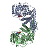
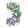
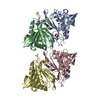
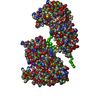
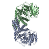
| Title | Structural definition of a unique neutralization epitope on the receptor-binding domain of MERS-CoV spike glycoprotein | ||||||
 Components Components |
| ||||||
 Keywords Keywords | VIRAL PROTEIN / Antibody | ||||||
| Function / homology |  Function and homology information Function and homology informationpositive regulation of viral entry into host cell / membrane fusion / host cell endoplasmic reticulum-Golgi intermediate compartment membrane / receptor-mediated virion attachment to host cell / endocytosis involved in viral entry into host cell / fusion of virus membrane with host plasma membrane / fusion of virus membrane with host endosome membrane / viral envelope / host cell plasma membrane / virion membrane / membrane Similarity search - Function | ||||||
| Biological species |   Middle East respiratory syndrome coronavirus Middle East respiratory syndrome coronavirus Homo sapiens (human) Homo sapiens (human) | ||||||
| Method |  X-RAY DIFFRACTION / SYNCHROTRON / MOLECULAR REPLACEMENT / Resolution: 4.482 Å X-RAY DIFFRACTION / SYNCHROTRON / MOLECULAR REPLACEMENT / Resolution: 4.482 Å | ||||||
 Authors Authors | Zhang, S. / Wang, X. | ||||||
| Funding support |  China, 1items China, 1items
| ||||||
 Citation Citation | Journal: Cell Rep / Year: 2018 Title: Structural Definition of a Unique Neutralization Epitope on the Receptor-Binding Domain of MERS-CoV Spike Glycoprotein Authors: Zhang, S. / Zhou, P. / Wang, P. / Li, Y. / Jiang, L. / Jia, W. / Wang, H. / Fan, A. / Wang, D. / Shi, X. / Fang, X. / Hammel, M. / Wang, S. / Wang, X. / Zhang, L. | ||||||
| History |
|
- Structure visualization
| Structure viewer | Molecule: MolmilJmol/JSmol |
|---|
- Downloads & links
-Download
| PDBx/mmCIF format | 5zxv.cif.gz | 494.8 KB | Display | PDBx/mmCIF format |
|---|---|---|---|---|
| PDB format | pdb5zxv.ent.gz | 417.3 KB | Display | PDB format |
| PDBx/mmJSON format | 5zxv.json.gz | Tree view | PDBx/mmJSON format | |
| Others |  Other downloads Other downloads |
-Validation report
| Arichive directory | https://data.pdbj.org/pub/pdb/validation_reports/zx/5zxvftp://data.pdbj.org/pub/pdb/validation_reports/zx/5zxv | HTTPS FTP |
|---|
-Related structure data
| Related structure data |  5yy5C  4l72S S: Starting model for refinement C: citing same article ( |
|---|---|
| Similar structure data |

-Links
 PDBj
PDBj

- Assembly
| Deposited unit | 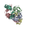
| ||||||||
|---|---|---|---|---|---|---|---|---|---|
| 1 | 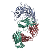
| ||||||||
| 2 | 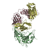
| ||||||||
| Unit cell |
|
-Components
| #1: Protein | Mass: 22929.961 Da / Num. of mol.: 2 Source method: isolated from a genetically manipulated source Source: (gene. exp.) Middle East respiratory syndrome coronavirusProduction host: Insect cell expression vector pTIE1 (others) References: UniProt: K9N5Q8*PLUS #2: Antibody | Mass: 22745.445 Da / Num. of mol.: 2 Source method: isolated from a genetically manipulated source Source: (gene. exp.) Homo sapiens (human) / Production host: Homo sapiens (human)#3: Antibody | Mass: 22724.953 Da / Num. of mol.: 2 Source method: isolated from a genetically manipulated source Source: (gene. exp.) Homo sapiens (human) / Production host: Homo sapiens (human)Has protein modification | Y | |
|---|
-Experimental details
-Experiment
| Experiment | Method: X-RAY DIFFRACTION / Number of used crystals: 1 |
|---|
- Sample preparation
| Crystal | Density Matthews: 3.13 Å3/Da / Density % sol: 60.76 % |
|---|---|
| Crystal grow | Temperature: 291.15 K / Method: vapor diffusion, sitting drop Details: 2%(v/v) tacsimate pH 5.0, 0.1M sodium citrate tribasic dihydrate pH 5.6, 16%(w/v) polyethylene glycol 3,350 and 2M sodium thiocyanate |
-Data collection
| Diffraction | Mean temperature: 100 K |
|---|---|
| Diffraction source | Source: SYNCHROTRON / Site: SSRF / Beamline: BL17U1 / Wavelength: 0.987 Å |
| Detector | Type: ADSC QUANTUM 315r / Detector: CCD / Date: May 15, 2016 |
| Radiation | Protocol: SINGLE WAVELENGTH / Monochromatic (M) / Laue (L): M / Scattering type: x-ray |
| Radiation wavelength | Wavelength: 0.987 Å / Relative weight: 1 |
| Reflection | Resolution: 4.482→50 Å / Num. obs: 9351 / % possible obs: 96.8 % / Redundancy: 3.1 % / Rmerge(I) obs: 0.108 / Net I/σ(I): 8.4 |
| Reflection shell | Resolution: 4.5→4.6 Å / Redundancy: 3.1 % / Num. unique obs: 9351 / CC1/2: 0.541 / % possible all: 97.8 |
- Processing
| Software |
| ||||||||||||||||||||||||||||||||||||||||
|---|---|---|---|---|---|---|---|---|---|---|---|---|---|---|---|---|---|---|---|---|---|---|---|---|---|---|---|---|---|---|---|---|---|---|---|---|---|---|---|---|---|
| Refinement | Method to determine structure: MOLECULAR REPLACEMENT Starting model: 4L72 Resolution: 4.482→35.438 Å / SU ML: 0.44 / Cross valid method: FREE R-VALUE / σ(F): 1.96 / Phase error: 52.66
| ||||||||||||||||||||||||||||||||||||||||
| Solvent computation | Shrinkage radii: 0.9 Å / VDW probe radii: 1.11 Å | ||||||||||||||||||||||||||||||||||||||||
| Refinement step | Cycle: LAST / Resolution: 4.482→35.438 Å
| ||||||||||||||||||||||||||||||||||||||||
| Refine LS restraints |
| ||||||||||||||||||||||||||||||||||||||||
| LS refinement shell |
| ||||||||||||||||||||||||||||||||||||||||
| Refinement TLS params. | Method: refined / Origin x: -37.7227 Å / Origin y: 91.9355 Å / Origin z: -3.3713 Å
| ||||||||||||||||||||||||||||||||||||||||
| Refinement TLS group | Selection details: all |
