 Movie
Movie Controller


+ Open data

- Basic information
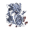
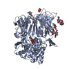
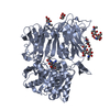
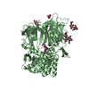
| Entry | Database: PDB / ID: 4l72 | |||||||||
|---|---|---|---|---|---|---|---|---|---|---|
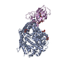
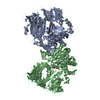
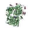
| Title | Crystal structure of MERS-CoV complexed with human DPP4 | |||||||||
 Components Components |
| |||||||||
 Keywords Keywords | HYDROLASE/VIRAL PROTEIN / alpha/beta hydrolase beta-propeller / Glycolation / virus receptor-binding domain / HYDROLASE-VIRAL PROTEIN complex | |||||||||
| Function / homology |  Function and homology information Function and homology informationglucagon processing / regulation of cell-cell adhesion mediated by integrin / negative regulation of neutrophil chemotaxis / Synthesis, secretion, and inactivation of Glucose-dependent Insulinotropic Polypeptide (GIP) / dipeptidyl-peptidase IV / negative regulation of extracellular matrix disassembly / chemorepellent activity / intercellular canaliculus / psychomotor behavior / dipeptidyl-peptidase activity ...glucagon processing / regulation of cell-cell adhesion mediated by integrin / negative regulation of neutrophil chemotaxis / Synthesis, secretion, and inactivation of Glucose-dependent Insulinotropic Polypeptide (GIP) / dipeptidyl-peptidase IV / negative regulation of extracellular matrix disassembly / chemorepellent activity / intercellular canaliculus / psychomotor behavior / dipeptidyl-peptidase activity / peptide hormone processing / lamellipodium membrane / behavioral fear response / locomotory exploration behavior / aminopeptidase activity / endothelial cell migration / endocytic vesicle / T cell costimulation / receptor-mediated endocytosis of virus by host cell / serine-type peptidase activity / T cell activation / Synthesis, secretion, and inactivation of Glucagon-like Peptide-1 (GLP-1) / lamellipodium / virus receptor activity / protease binding / membrane fusion / response to hypoxia / host cell endoplasmic reticulum-Golgi intermediate compartment membrane / receptor-mediated virion attachment to host cell / cell adhesion / apical plasma membrane / endocytosis involved in viral entry into host cell / membrane raft / serine-type endopeptidase activity / fusion of virus membrane with host plasma membrane / signaling receptor binding / lysosomal membrane / focal adhesion / fusion of virus membrane with host endosome membrane / positive regulation of cell population proliferation / viral envelope / symbiont entry into host cell / host cell plasma membrane / virion membrane / cell surface / protein homodimerization activity / proteolysis / extracellular exosome / extracellular region / membrane / identical protein binding / plasma membrane Similarity search - Function | |||||||||
| Biological species |  Homo sapiens (human) Homo sapiens (human) 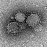 Middle East respiratory syndrome coronavirus Middle East respiratory syndrome coronavirus | |||||||||
| Method |  X-RAY DIFFRACTION / SYNCHROTRON / MOLECULAR REPLACEMENT / Resolution: 3.005 Å X-RAY DIFFRACTION / SYNCHROTRON / MOLECULAR REPLACEMENT / Resolution: 3.005 Å | |||||||||
 Authors Authors | Wang, X.Q. / Wang, N.S. | |||||||||
 Citation Citation | Journal: Cell Res. / Year: 2013 Title: Structure of MERS-CoV spike receptor-binding domain complexed with human receptor DPP4 Authors: Wang, N. / Shi, X. / Jiang, L. / Zhang, S. / Wang, D. / Tong, P. / Guo, D. / Fu, L. / Cui, Y. / Liu, X. / Arledge, K.C. / Chen, Y.H. / Zhang, L. / Wang, X. | |||||||||
| History |
|
- Structure visualization
| Structure viewer | Molecule: MolmilJmol/JSmol |
|---|
- Downloads & links
-Download
| PDBx/mmCIF format | 4l72.cif.gz | 379.8 KB | Display | PDBx/mmCIF format |
|---|---|---|---|---|
| PDB format | pdb4l72.ent.gz | 311 KB | Display | PDB format |
| PDBx/mmJSON format | 4l72.json.gz | Tree view | PDBx/mmJSON format | |
| Others |  Other downloads Other downloads |
-Validation report
| Arichive directory | https://data.pdbj.org/pub/pdb/validation_reports/l7/4l72ftp://data.pdbj.org/pub/pdb/validation_reports/l7/4l72 | HTTPS FTP |
|---|
-Related structure data


-Links
 PDBj
PDBj


- Assembly
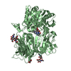
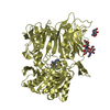
| Deposited unit | 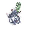
| ||||||||
|---|---|---|---|---|---|---|---|---|---|
| 1 | 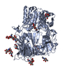
| ||||||||
| 2 | 
| ||||||||
| 3 | 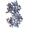
| ||||||||
| Unit cell |
|
-Components
| #1: Protein | Mass: 84462.617 Da / Num. of mol.: 1 / Fragment: UNP residues 39-766 Source method: isolated from a genetically manipulated source Source: (gene. exp.) Homo sapiens (human) / Gene: DPP4, ADCP2, CD26 / Production host:   Spodoptera frugiperda (fall armyworm) / References: UniProt: P27487, dipeptidyl-peptidase IV Spodoptera frugiperda (fall armyworm) / References: UniProt: P27487, dipeptidyl-peptidase IV | ||||||
|---|---|---|---|---|---|---|---|
| #2: Protein | Mass: 22491.373 Da / Num. of mol.: 1 Source method: isolated from a genetically manipulated source Source: (gene. exp.) Middle East respiratory syndrome coronavirusProduction host: Spodoptera frugiperda (fall armyworm) / References: UniProt: R9UQ53*PLUS | ||||||
| #3: Polysaccharide | Source method: isolated from a genetically manipulated source #4: Polysaccharide | beta-D-mannopyranose-(1-4)-2-acetamido-2-deoxy-beta-D-glucopyranose-(1-4)-2-acetamido-2-deoxy-beta- ...beta-D-mannopyranose-(1-4)-2-acetamido-2-deoxy-beta-D-glucopyranose-(1-4)-2-acetamido-2-deoxy-beta-D-glucopyranose | Source method: isolated from a genetically manipulated source #5: Sugar | ChemComp-NAG /   Type: D-saccharide, beta linking / Mass: 221.208 Da / Num. of mol.: 5 Type: D-saccharide, beta linking / Mass: 221.208 Da / Num. of mol.: 5Source method: isolated from a genetically manipulated source Formula: C8H15NO6 Has protein modification | Y | |
-Experimental details
-Experiment
| Experiment | Method: X-RAY DIFFRACTION / Number of used crystals: 1 |
|---|
- Sample preparation
| Crystal | Density Matthews: 4.36 Å3/Da / Density % sol: 71.77 % |
|---|---|
| Crystal grow | Temperature: 291 K / Method: vapor diffusion, sitting drop / pH: 7.2 Details: 20% PEG1500, pH 7.2, VAPOR DIFFUSION, SITTING DROP, temperature 291K |
-Data collection
| Diffraction | Mean temperature: 100 K |
|---|---|
| Diffraction source | Source: SYNCHROTRON / Site: SSRF  / Beamline: BL17U / Wavelength: 0.97 Å / Beamline: BL17U / Wavelength: 0.97 Å |
| Detector | Type: RIGAKU / Detector: IMAGE PLATE / Date: Apr 5, 2013 |
| Radiation | Monochromator: GRAPHITE / Protocol: SINGLE WAVELENGTH / Monochromatic (M) / Laue (L): M / Scattering type: x-ray |
| Radiation wavelength | Wavelength: 0.97 Å / Relative weight: 1 |
| Reflection | Resolution: 3→48.9 Å / Num. all: 39446 / Num. obs: 39446 / % possible obs: 99.6 % / Observed criterion σ(F): 1.34 / Observed criterion σ(I): 7.2 |
| Reflection shell | Resolution: 3→48.9 Å / % possible all: 99.6 |
- Processing
| Software |
| ||||||||||||||||||||||||||||||||||||||||||||||||||||||||||||||||||||||||||||||||||||||||||
|---|---|---|---|---|---|---|---|---|---|---|---|---|---|---|---|---|---|---|---|---|---|---|---|---|---|---|---|---|---|---|---|---|---|---|---|---|---|---|---|---|---|---|---|---|---|---|---|---|---|---|---|---|---|---|---|---|---|---|---|---|---|---|---|---|---|---|---|---|---|---|---|---|---|---|---|---|---|---|---|---|---|---|---|---|---|---|---|---|---|---|---|
| Refinement | Method to determine structure: MOLECULAR REPLACEMENT Starting model: 2G63, 2AJF Resolution: 3.005→48.9 Å / SU ML: 0.34 / σ(F): 1.34 / Phase error: 23.95 / Stereochemistry target values: ML
| ||||||||||||||||||||||||||||||||||||||||||||||||||||||||||||||||||||||||||||||||||||||||||
| Solvent computation | Shrinkage radii: 0.9 Å / VDW probe radii: 1.11 Å / Solvent model: FLAT BULK SOLVENT MODEL | ||||||||||||||||||||||||||||||||||||||||||||||||||||||||||||||||||||||||||||||||||||||||||
| Refinement step | Cycle: LAST / Resolution: 3.005→48.9 Å
| ||||||||||||||||||||||||||||||||||||||||||||||||||||||||||||||||||||||||||||||||||||||||||
| Refine LS restraints |
| ||||||||||||||||||||||||||||||||||||||||||||||||||||||||||||||||||||||||||||||||||||||||||
| LS refinement shell | Refine-ID: X-RAY DIFFRACTION / Total num. of bins used: 14
| ||||||||||||||||||||||||||||||||||||||||||||||||||||||||||||||||||||||||||||||||||||||||||
| Refinement TLS params. | Method: refined / Refine-ID: X-RAY DIFFRACTION
| ||||||||||||||||||||||||||||||||||||||||||||||||||||||||||||||||||||||||||||||||||||||||||
| Refinement TLS group |
|
