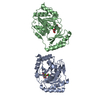
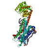
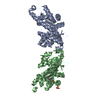
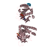
Entry Database : PDB / ID : 5zxiTitle Co-crystal structure of an Inhibitor in complex with human PPARdelta LBD Peroxisome proliferator-activated receptor delta Keywords / / Function / homology Function Domain/homology Component
/ / / / / / / / / / / / / / / / / / / / / / / / / / / / / / / / / / / / / / / / / / / / / / / / / / / / / / / / / / / / / / / / / / / / / / / / / / / / / / / / / / / / / / / / / / / / / / / / / / / / / / / / / / / / / / / / / / / / Biological species Homo sapiens (human)Method / / / Resolution : 2.1 Å Authors Rani, S.T. / Laxminarasimhan, A. / Senaiar, R.S. / Krishnamurthy, N. Journal : ACS Med Chem Lett / Year : 2018Title : Selective PPAR delta Modulators Improve Mitochondrial Function: Potential Treatment for Duchenne Muscular Dystrophy (DMD).Authors: Lagu, B. / Kluge, A.F. / Tozzo, E. / Fredenburg, R. / Bell, E.L. / Goddeeris, M.M. / Dwyer, P. / Basinski, A. / Senaiar, R.S. / Jaleel, M. / Tiwari, N.K. / Panigrahi, S.K. / Krishnamurthy, N. ... Authors : Lagu, B. / Kluge, A.F. / Tozzo, E. / Fredenburg, R. / Bell, E.L. / Goddeeris, M.M. / Dwyer, P. / Basinski, A. / Senaiar, R.S. / Jaleel, M. / Tiwari, N.K. / Panigrahi, S.K. / Krishnamurthy, N.R. / Takahashi, T. / Patane, M.A. History Deposition May 21, 2018 Deposition site / Processing site Revision 1.0 Apr 3, 2019 Provider / Type Revision 1.1 Mar 27, 2024 Group / Database references / Category / chem_comp_bond / database_2Item / _database_2.pdbx_database_accession
Show all Show less
 Movie
Movie Controller

 Yorodumi
 Open data
 Basic information
 Components
 Keywords
 Function and homology information
 Homo sapiens (human)
 X-RAY DIFFRACTION /
  Authors
 Citation
 Structure visualization
 Downloads & links
 Other downloads
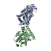


 PDBj
PDBj
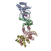
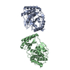
 Assembly
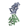

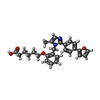
 Mass: 444.522 Da / Num. of mol.: 2 / Source method: obtained synthetically / Formula: C27H28N2O4 / Feature type: SUBJECT OF INVESTIGATION
 Mass: 18.015 Da / Num. of mol.: 151 / Source method: isolated from a natural source / Formula: H2O
 Sample preparation
 / Beamline: MX2 / Wavelength: 1 Å
 Processing