 Movie
Movie Controller

[English] 日本語
 Yorodumi
- PDB-5zwa: Crystal structure of Pyridoxal kinase (PdxK) from Salmonella typh... -
+ Open data

- Basic information
| Entry | Database: PDB / ID: 5zwa | |||||||||
|---|---|---|---|---|---|---|---|---|---|---|
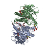
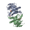
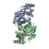
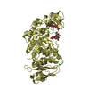
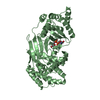
| Title | Crystal structure of Pyridoxal kinase (PdxK) from Salmonella typhimurium in complex with ADP, PL-linked to Lys233 via Schiff base in protomer A and the product (PLP) in protomer B | |||||||||
 Components Components | (Pyridoxine/pyridoxal/pyridoxamine ...) x 2 | |||||||||
 Keywords Keywords | TRANSFERASE / Pyridoxal kinase / bound phosphate / Salmonella typhimurium | |||||||||
| Function / homology |  Function and homology information Function and homology informationhydroxymethylpyrimidine kinase activity / pyridoxal 5'-phosphate salvage / pyridoxal kinase / pyridoxal kinase activity / magnesium ion binding / zinc ion binding / ATP binding / cytosol Similarity search - Function | |||||||||
| Biological species |  Salmonella choleraesuis (bacteria) Salmonella choleraesuis (bacteria) | |||||||||
| Method |  X-RAY DIFFRACTION / MOLECULAR REPLACEMENT / Resolution: 2.45 Å X-RAY DIFFRACTION / MOLECULAR REPLACEMENT / Resolution: 2.45 Å | |||||||||
 Authors Authors | Deka, G. / Benazir, J.F. / Kalyani, J.N. / Savithri, H.S. / Murthy, M.R.N. | |||||||||
| Funding support |  India, 2items India, 2items
| |||||||||
 Citation Citation | Journal: Febs J. / Year: 2019 Title: Structural and functional studies on Salmonella typhimurium pyridoxal kinase: the first structural evidence for the formation of Schiff base with the substrate. Authors: Deka, G. / Kalyani, J.N. / Jahangir, F.B. / Sabharwal, P. / Savithri, H.S. / Murthy, M.R.N. | |||||||||
| History |
|
- Structure visualization
| Structure viewer | Molecule: MolmilJmol/JSmol |
|---|
- Downloads & links
-Download
| PDBx/mmCIF format | 5zwa.cif.gz | 127.2 KB | Display | PDBx/mmCIF format |
|---|---|---|---|---|
| PDB format | pdb5zwa.ent.gz | 94.2 KB | Display | PDB format |
| PDBx/mmJSON format | 5zwa.json.gz | Tree view | PDBx/mmJSON format | |
| Others |  Other downloads Other downloads |
-Validation report
| Arichive directory | https://data.pdbj.org/pub/pdb/validation_reports/zw/5zwaftp://data.pdbj.org/pub/pdb/validation_reports/zw/5zwa | HTTPS FTP |
|---|
-Related structure data
| Related structure data |  5zw9SC  5zwbC S: Starting model for refinement C: citing same article ( |
|---|---|
| Similar structure data |


-Links
 PDBj
PDBj- Assembly
| Deposited unit | 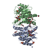
| ||||||||||||||||||
|---|---|---|---|---|---|---|---|---|---|---|---|---|---|---|---|---|---|---|---|
| 1 |
| ||||||||||||||||||
| Unit cell |
| ||||||||||||||||||
| Noncrystallographic symmetry (NCS) | NCS domain:
NCS domain segments: Component-ID: _ / Ens-ID: 1 / Beg auth comp-ID: ARG / Beg label comp-ID: ARG / End auth comp-ID: PRO / End label comp-ID: PRO / Refine code: _ / Auth seq-ID: 17 - 283 / Label seq-ID: 17 - 283
|
-Components
-Pyridoxine/pyridoxal/pyridoxamine ... , 2 types, 2 molecules AB
| #1: Protein | Mass: 32287.844 Da / Num. of mol.: 1 Source method: isolated from a genetically manipulated source Source: (gene. exp.) Salmonella choleraesuis (bacteria) / Gene: pdxK / Production host: References: UniProt: A0A0M0PWM4, UniProt: A0A0F7J8S0*PLUS, pyridoxal kinase |
|---|---|
| #2: Protein | Mass: 32139.703 Da / Num. of mol.: 1 Source method: isolated from a genetically manipulated source Source: (gene. exp.) Salmonella choleraesuis (bacteria) / Gene: pdxK / Plasmid: pET-22b(+) / Production host: References: UniProt: A0A0M0PWM4, UniProt: A0A0F7J8S0*PLUS, pyridoxal kinase |
-Non-polymers , 8 types, 282 molecules 


| #3: Chemical |  Mass: 96.063 Da / Num. of mol.: 3 / Source method: obtained synthetically / Formula: SO4 Mass: 96.063 Da / Num. of mol.: 3 / Source method: obtained synthetically / Formula: SO4#4: Chemical | ChemComp-EDO /  Mass: 62.068 Da / Num. of mol.: 15 / Source method: obtained synthetically / Formula: C2H6O2 Mass: 62.068 Da / Num. of mol.: 15 / Source method: obtained synthetically / Formula: C2H6O2#5: Chemical | ChemComp-ADP / |  Mass: 427.201 Da / Num. of mol.: 1 / Source method: obtained synthetically / Formula: C10H15N5O10P2 / Comment: ADP, energy-carrying molecule*YM Mass: 427.201 Da / Num. of mol.: 1 / Source method: obtained synthetically / Formula: C10H15N5O10P2 / Comment: ADP, energy-carrying molecule*YM#6: Chemical |  Mass: 24.305 Da / Num. of mol.: 2 / Source method: obtained synthetically / Formula: Mg Mass: 24.305 Da / Num. of mol.: 2 / Source method: obtained synthetically / Formula: Mg#7: Chemical |  Mass: 92.094 Da / Num. of mol.: 2 / Source method: obtained synthetically / Formula: C3H8O3 Mass: 92.094 Da / Num. of mol.: 2 / Source method: obtained synthetically / Formula: C3H8O3#8: Chemical | ChemComp-PLP / |  Mass: 247.142 Da / Num. of mol.: 1 / Source method: obtained synthetically / Formula: C8H10NO6P Mass: 247.142 Da / Num. of mol.: 1 / Source method: obtained synthetically / Formula: C8H10NO6P#9: Chemical | ChemComp-PO4 / |  Mass: 94.971 Da / Num. of mol.: 1 / Source method: obtained synthetically / Formula: PO4 Mass: 94.971 Da / Num. of mol.: 1 / Source method: obtained synthetically / Formula: PO4#10: Water | ChemComp-HOH / | Mass: 18.015 Da / Num. of mol.: 257 / Source method: isolated from a natural source / Formula: H2O |
|---|
-Experimental details
-Experiment
| Experiment | Method: X-RAY DIFFRACTION / Number of used crystals: 1 |
|---|
- Sample preparation
| Crystal | Density Matthews: 2.42 Å3/Da / Density % sol: 49.12 % |
|---|---|
| Crystal grow | Temperature: 293 K / Method: batch mode / pH: 8.5 / Details: 50% PEG 4000, 10% Glycerol, 100 mM Tris |
-Data collection
| Diffraction | Mean temperature: 100 K |
|---|---|
| Diffraction source | Source: ROTATING ANODE / Type: RIGAKU / Wavelength: 1.5417 Å |
| Detector | Type: MARMOSAIC 225 mm CCD / Detector: CCD / Date: Jan 26, 2013 |
| Radiation | Protocol: SINGLE WAVELENGTH / Monochromatic (M) / Laue (L): M / Scattering type: x-ray |
| Radiation wavelength | Wavelength: 1.5417 Å / Relative weight: 1 |
| Reflection | Resolution: 2.45→62.23 Å / Num. obs: 23479 / % possible obs: 94.3 % / Redundancy: 14.9 % / Biso Wilson estimate: 22.9 Å2 / Rmerge(I) obs: 0.13 / Rpim(I) all: 0.03 / Rrim(I) all: 0.14 / Net I/σ(I): 15.7 |
| Reflection shell | Resolution: 2.45→2.58 Å / Redundancy: 15.6 % / Rmerge(I) obs: 0.49 / Mean I/σ(I) obs: 5.5 / Num. unique obs: 3297 / Rpim(I) all: 0.11 / Rrim(I) all: 0.5 / % possible all: 93 |
- Processing
| Software |
| ||||||||||||||||||||||||||||||||||||||||||||||||||||||||||||||||||||||||||||||||||||||||||||||||||||||||||||||||||||||||||||||||||||||||||||||||||||||||||||||||||||||||||||||||||||||
|---|---|---|---|---|---|---|---|---|---|---|---|---|---|---|---|---|---|---|---|---|---|---|---|---|---|---|---|---|---|---|---|---|---|---|---|---|---|---|---|---|---|---|---|---|---|---|---|---|---|---|---|---|---|---|---|---|---|---|---|---|---|---|---|---|---|---|---|---|---|---|---|---|---|---|---|---|---|---|---|---|---|---|---|---|---|---|---|---|---|---|---|---|---|---|---|---|---|---|---|---|---|---|---|---|---|---|---|---|---|---|---|---|---|---|---|---|---|---|---|---|---|---|---|---|---|---|---|---|---|---|---|---|---|---|---|---|---|---|---|---|---|---|---|---|---|---|---|---|---|---|---|---|---|---|---|---|---|---|---|---|---|---|---|---|---|---|---|---|---|---|---|---|---|---|---|---|---|---|---|---|---|---|---|
| Refinement | Method to determine structure: MOLECULAR REPLACEMENT Starting model: 5ZW9 Resolution: 2.45→62.23 Å / Cor.coef. Fo:Fc: 0.918 / Cor.coef. Fo:Fc free: 0.901 / SU B: 7.827 / SU ML: 0.178 / Cross valid method: THROUGHOUT / ESU R: 0.527 / ESU R Free: 0.264 / Details: HYDROGENS HAVE BEEN ADDED IN THE RIDING POSITIONS
| ||||||||||||||||||||||||||||||||||||||||||||||||||||||||||||||||||||||||||||||||||||||||||||||||||||||||||||||||||||||||||||||||||||||||||||||||||||||||||||||||||||||||||||||||||||||
| Solvent computation | Ion probe radii: 0.8 Å / Shrinkage radii: 0.8 Å / VDW probe radii: 1.2 Å | ||||||||||||||||||||||||||||||||||||||||||||||||||||||||||||||||||||||||||||||||||||||||||||||||||||||||||||||||||||||||||||||||||||||||||||||||||||||||||||||||||||||||||||||||||||||
| Displacement parameters | Biso mean: 26.782 Å2
| ||||||||||||||||||||||||||||||||||||||||||||||||||||||||||||||||||||||||||||||||||||||||||||||||||||||||||||||||||||||||||||||||||||||||||||||||||||||||||||||||||||||||||||||||||||||
| Refinement step | Cycle: 1 / Resolution: 2.45→62.23 Å
| ||||||||||||||||||||||||||||||||||||||||||||||||||||||||||||||||||||||||||||||||||||||||||||||||||||||||||||||||||||||||||||||||||||||||||||||||||||||||||||||||||||||||||||||||||||||
| Refine LS restraints |
|