 Movie
Movie Controller


+ Open data

- Basic information
| Entry | Database: PDB / ID: 5zm2 | ||||||
|---|---|---|---|---|---|---|---|
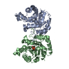
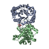
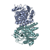
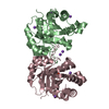
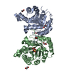
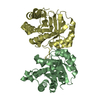
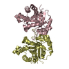
| Title | Fe(II)/(alpha)ketoglutarate-dependent dioxygenase AndA | ||||||
 Components Components | Dioxygenase andA | ||||||
 Keywords Keywords | OXIDOREDUCTASE | ||||||
| Function / homology |  Function and homology information Function and homology informationterpenoid biosynthetic process / Oxidoreductases; Acting on paired donors, with incorporation or reduction of molecular oxygen; With 2-oxoglutarate as one donor, and incorporation of one atom of oxygen into each donor / dioxygenase activity / metal ion binding Similarity search - Function | ||||||
| Biological species |  | ||||||
| Method |  X-RAY DIFFRACTION / SYNCHROTRON / MOLECULAR REPLACEMENT / Resolution: 2.5 Å X-RAY DIFFRACTION / SYNCHROTRON / MOLECULAR REPLACEMENT / Resolution: 2.5 Å | ||||||
 Authors Authors | Nakashima, Y. / Senda, T. | ||||||
 Citation Citation | Journal: J. Am. Chem. Soc. / Year: 2018 Title: Structural and Computational Bases for Dramatic Skeletal Rearrangement in Anditomin Biosynthesis. Authors: Nakashima, Y. / Mitsuhashi, T. / Matsuda, Y. / Senda, M. / Sato, H. / Yamazaki, M. / Uchiyama, M. / Senda, T. / Abe, I. | ||||||
| History |
|
- Structure visualization
| Structure viewer | Molecule: MolmilJmol/JSmol |
|---|
- Downloads & links
-Download
| PDBx/mmCIF format | 5zm2.cif.gz | 216.2 KB | Display | PDBx/mmCIF format |
|---|---|---|---|---|
| PDB format | pdb5zm2.ent.gz | 169.9 KB | Display | PDB format |
| PDBx/mmJSON format | 5zm2.json.gz | Tree view | PDBx/mmJSON format | |
| Others |  Other downloads Other downloads |
-Validation report
| Arichive directory | https://data.pdbj.org/pub/pdb/validation_reports/zm/5zm2ftp://data.pdbj.org/pub/pdb/validation_reports/zm/5zm2 | HTTPS FTP |
|---|
-Related structure data
| Related structure data | 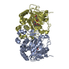 5zm3C 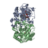 5zm4C 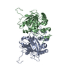 5ybmS S: Starting model for refinement C: citing same article ( |
|---|---|
| Similar structure data |


-Links
 PDBj
PDBj- Assembly
| Deposited unit | 
| ||||||||
|---|---|---|---|---|---|---|---|---|---|
| 1 | 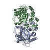
| ||||||||
| 2 | 
| ||||||||
| Unit cell |
|
-Components
| #1: Protein | Mass: 33854.402 Da / Num. of mol.: 4 Source method: isolated from a genetically manipulated source Source: (gene. exp.)  References: UniProt: A0A097ZPD5, Oxidoreductases; Acting on paired donors, with incorporation or reduction of molecular oxygen; With 2-oxoglutarate as one donor, and incorporation of one atom of oxygen into each donor #2: Water | ChemComp-HOH / |  Mass: 18.015 Da / Num. of mol.: 245 / Source method: isolated from a natural source / Formula: H2O Mass: 18.015 Da / Num. of mol.: 245 / Source method: isolated from a natural source / Formula: H2O |
|---|
-Experimental details
-Experiment
| Experiment | Method: X-RAY DIFFRACTION / Number of used crystals: 1 |
|---|
- Sample preparation
| Crystal | Density Matthews: 2.75 Å3/Da / Density % sol: 55.31 % |
|---|---|
| Crystal grow | Temperature: 293 K / Method: vapor diffusion, sitting drop / Details: 30% (w/v) PEG 3350, 200mM sodium tartrate, 20% DMF |
-Data collection
| Diffraction | Mean temperature: 95 K |
|---|---|
| Diffraction source | Source: SYNCHROTRON / Site: Photon Factory  / Beamline: BL-1A / Wavelength: 1.1 Å / Beamline: BL-1A / Wavelength: 1.1 Å |
| Detector | Type: DECTRIS EIGER X 4M / Detector: PIXEL / Date: Apr 29, 2017 |
| Radiation | Protocol: SINGLE WAVELENGTH / Monochromatic (M) / Laue (L): M / Scattering type: x-ray |
| Radiation wavelength | Wavelength: 1.1 Å / Relative weight: 1 |
| Reflection | Resolution: 2.5→47.55 Å / Num. obs: 48298 / % possible obs: 96.7 % / Redundancy: 3.4 % / Rmerge(I) obs: 0.149 / Net I/σ(I): 6.6 |
| Reflection shell | Resolution: 2.5→2.55 Å / Rmerge(I) obs: 0.511 / Num. unique obs: 4442 |
- Processing
| Software |
| ||||||||||||||||||||||||||||||||||||||||||||||||||||||||||||||||||||||||||||||||||||||||||||||||||||||||||||||||||||||||||||||
|---|---|---|---|---|---|---|---|---|---|---|---|---|---|---|---|---|---|---|---|---|---|---|---|---|---|---|---|---|---|---|---|---|---|---|---|---|---|---|---|---|---|---|---|---|---|---|---|---|---|---|---|---|---|---|---|---|---|---|---|---|---|---|---|---|---|---|---|---|---|---|---|---|---|---|---|---|---|---|---|---|---|---|---|---|---|---|---|---|---|---|---|---|---|---|---|---|---|---|---|---|---|---|---|---|---|---|---|---|---|---|---|---|---|---|---|---|---|---|---|---|---|---|---|---|---|---|---|
| Refinement | Method to determine structure: MOLECULAR REPLACEMENT Starting model: 5YBM Resolution: 2.5→47.078 Å / Cross valid method: FREE R-VALUE / σ(F): 1.97 / Phase error: 27.55
| ||||||||||||||||||||||||||||||||||||||||||||||||||||||||||||||||||||||||||||||||||||||||||||||||||||||||||||||||||||||||||||||
| Solvent computation | Shrinkage radii: 0.9 Å / VDW probe radii: 1.11 Å | ||||||||||||||||||||||||||||||||||||||||||||||||||||||||||||||||||||||||||||||||||||||||||||||||||||||||||||||||||||||||||||||
| Refinement step | Cycle: LAST / Resolution: 2.5→47.078 Å
| ||||||||||||||||||||||||||||||||||||||||||||||||||||||||||||||||||||||||||||||||||||||||||||||||||||||||||||||||||||||||||||||
| Refine LS restraints |
| ||||||||||||||||||||||||||||||||||||||||||||||||||||||||||||||||||||||||||||||||||||||||||||||||||||||||||||||||||||||||||||||
| LS refinement shell |
|
