 Movie
Movie Controller

[English] 日本語
 Yorodumi
- PDB-5zl2: Crystal structure of Bourbon virus envelope glycoprotein at pH8.0 -
+ Open data

- Basic information
| Entry | Database: PDB / ID: 5zl2 | |||||||||
|---|---|---|---|---|---|---|---|---|---|---|
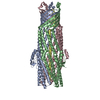
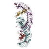
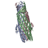
| Title | Crystal structure of Bourbon virus envelope glycoprotein at pH8.0 | |||||||||
 Components Components | Envelope glycoprotein | |||||||||
 Keywords Keywords | VIRAL PROTEIN / BOUV / glycoprotein | |||||||||
| Function / homology | Baculovirus Gp64, envelope glycoprotein / Baculovirus gp64 envelope glycoprotein / symbiont-mediated perturbation of host process / viral envelope / virion membrane / Envelope glycoprotein Function and homology information Function and homology information | |||||||||
| Biological species |  Bourbon virus Bourbon virus | |||||||||
| Method |  X-RAY DIFFRACTION / SYNCHROTRON / MOLECULAR REPLACEMENT / Resolution: 2.703 Å X-RAY DIFFRACTION / SYNCHROTRON / MOLECULAR REPLACEMENT / Resolution: 2.703 Å | |||||||||
 Authors Authors | Qi, J.X. / Peng, R.C. / Wu, Y. / Gao, F. | |||||||||
| Funding support |  China, 2items China, 2items
| |||||||||
 Citation Citation | Journal: To Be Published Title: The postfusion structure of human-infecting Bourbon virus envelope glycoprotein implicates the host adaptation properties of thogotoviruses Authors: Bai, C.Z. / Qi, J.X. / Wu, Y. / Wang, X.J. / Gao, G.F. / Peng, R.C. / Gao, F. | |||||||||
| History |
|
- Structure visualization
| Structure viewer | Molecule: MolmilJmol/JSmol |
|---|
- Downloads & links
-Download
| PDBx/mmCIF format | 5zl2.cif.gz | 447.4 KB | Display | PDBx/mmCIF format |
|---|---|---|---|---|
| PDB format | pdb5zl2.ent.gz | 367.6 KB | Display | PDB format |
| PDBx/mmJSON format | 5zl2.json.gz | Tree view | PDBx/mmJSON format | |
| Others |  Other downloads Other downloads |
-Validation report
| Arichive directory | https://data.pdbj.org/pub/pdb/validation_reports/zl/5zl2ftp://data.pdbj.org/pub/pdb/validation_reports/zl/5zl2 | HTTPS FTP |
|---|
-Related structure data
| Related structure data | |
|---|---|
| Similar structure data |


-Links
 PDBj
PDBj- Assembly
| Deposited unit | 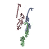
| ||||||||
|---|---|---|---|---|---|---|---|---|---|
| 1 | 
| ||||||||
| 2 | 
| ||||||||
| 3 | 
| ||||||||
| Unit cell |
|
-Components
| #1: Protein | Mass: 52931.625 Da / Num. of mol.: 3 / Fragment: UNP residues 19-485 Source method: isolated from a genetically manipulated source Source: (gene. exp.) Bourbon virus / Production host: Baculovirus expression vector pFastBac1-HM / References: UniProt: A0A140H4W8#2: Water | ChemComp-HOH / |  Mass: 18.015 Da / Num. of mol.: 116 / Source method: isolated from a natural source / Formula: H2O Mass: 18.015 Da / Num. of mol.: 116 / Source method: isolated from a natural source / Formula: H2OHas protein modification | Y | |
|---|
-Experimental details
-Experiment
| Experiment | Method: X-RAY DIFFRACTION / Number of used crystals: 1 |
|---|
- Sample preparation
| Crystal | Density Matthews: 2.56 Å3/Da / Density % sol: 51.88 % |
|---|---|
| Crystal grow | Temperature: 291 K / Method: vapor diffusion, sitting drop / pH: 8 Details: 0.1M Tris, pH 8.0 and 30%(v/v) polyethylene glycol 400 |
-Data collection
| Diffraction | Mean temperature: 100 K |
|---|---|
| Diffraction source | Source: SYNCHROTRON / Site: SSRF / Beamline: BL19U1 / Wavelength: 0.97917 Å |
| Detector | Type: DECTRIS PILATUS3 6M / Detector: PIXEL / Date: Mar 23, 2018 |
| Radiation | Protocol: SINGLE WAVELENGTH / Monochromatic (M) / Laue (L): M / Scattering type: x-ray |
| Radiation wavelength | Wavelength: 0.97917 Å / Relative weight: 1 |
| Reflection twin | Operator: -k,-h,-l / Fraction: 0.48 |
| Reflection | Resolution: 2.7→50 Å / Num. obs: 42840 / % possible obs: 99.4 % / Redundancy: 5.3 % / CC1/2: 0.961 / Rmerge(I) obs: 0.186 / Rpim(I) all: 0.085 / Net I/σ(I): 8.579 |
| Reflection shell | Resolution: 2.7→2.8 Å / Mean I/σ(I) obs: 1.09 / CC1/2: 0.662 / Rpim(I) all: 0.483 |
- Processing
| Software |
| |||||||||||||||||||||||||||||||||||||||||||||||||||||||||||||||||||||||||||||||||||||||||||
|---|---|---|---|---|---|---|---|---|---|---|---|---|---|---|---|---|---|---|---|---|---|---|---|---|---|---|---|---|---|---|---|---|---|---|---|---|---|---|---|---|---|---|---|---|---|---|---|---|---|---|---|---|---|---|---|---|---|---|---|---|---|---|---|---|---|---|---|---|---|---|---|---|---|---|---|---|---|---|---|---|---|---|---|---|---|---|---|---|---|---|---|---|
| Refinement | Method to determine structure: MOLECULAR REPLACEMENT / Resolution: 2.703→47.79 Å / Cross valid method: FREE R-VALUE / σ(F): 2 / Phase error: 24.16
| |||||||||||||||||||||||||||||||||||||||||||||||||||||||||||||||||||||||||||||||||||||||||||
| Solvent computation | Shrinkage radii: 0.9 Å / VDW probe radii: 1.11 Å | |||||||||||||||||||||||||||||||||||||||||||||||||||||||||||||||||||||||||||||||||||||||||||
| Refinement step | Cycle: LAST / Resolution: 2.703→47.79 Å
| |||||||||||||||||||||||||||||||||||||||||||||||||||||||||||||||||||||||||||||||||||||||||||
| Refine LS restraints |
| |||||||||||||||||||||||||||||||||||||||||||||||||||||||||||||||||||||||||||||||||||||||||||
| LS refinement shell |
| |||||||||||||||||||||||||||||||||||||||||||||||||||||||||||||||||||||||||||||||||||||||||||
| Refinement TLS params. | Method: refined / Origin x: 19.6296 Å / Origin y: 26.1858 Å / Origin z: -23.1632 Å
| |||||||||||||||||||||||||||||||||||||||||||||||||||||||||||||||||||||||||||||||||||||||||||
| Refinement TLS group | Selection details: all |
