- PDB-5zib: Crystal structure of human GnT-V luminal domain in apo form -
+
Open data
ID or keywords:
Loading...
-
Basic information
Entry
Database: PDB / ID: 5zib
Title
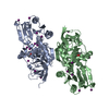
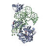
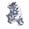
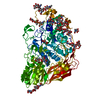
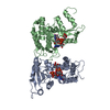
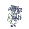
Crystal structure of human GnT-V luminal domain in apo form
Components
Alpha-1,6-mannosylglycoprotein 6-beta-N-acetylglucosaminyltransferase A
Keywords
SUGAR BINDING PROTEIN / Glycosyltransferase Cancer metastasis Luminal protein
Function / homology
Function and homology information
alpha-1,6-mannosyl-glycoprotein 6-beta-N-acetylglucosaminyltransferase / alpha-1,6-mannosylglycoprotein 6-beta-N-acetylglucosaminyltransferase activity / N-Glycan antennae elongation / positive regulation of receptor signaling pathway via STAT / negative regulation of protein tyrosine phosphatase activity / protein N-linked glycosylation via asparagine / protein N-linked glycosylation / protein phosphatase inhibitor activity / manganese ion binding / Maturation of spike protein ...alpha-1,6-mannosyl-glycoprotein 6-beta-N-acetylglucosaminyltransferase / alpha-1,6-mannosylglycoprotein 6-beta-N-acetylglucosaminyltransferase activity / N-Glycan antennae elongation / positive regulation of receptor signaling pathway via STAT / negative regulation of protein tyrosine phosphatase activity / protein N-linked glycosylation via asparagine / protein N-linked glycosylation / protein phosphatase inhibitor activity / manganese ion binding / Maturation of spike protein / viral protein processing / positive regulation of cell migration / Golgi membrane / Golgi apparatus / extracellular exosome / membrane Similarity search - Function
Glycosyltransferase family 18 / Domain of unknown function DUF4525 / Glycosyltransferase family 18 / Domain of unknown function (DUF4525) Similarity search - Domain/homology
In the structure databanks used in Yorodumi, some data are registered as the other names, "COVID-19 virus" and "2019-nCoV". Here are the details of the virus and the list of structure data.
Jan 31, 2019. EMDB accession codes are about to change! (news from PDBe EMDB page)
EMDB accession codes are about to change! (news from PDBe EMDB page)
The allocation of 4 digits for EMDB accession codes will soon come to an end. Whilst these codes will remain in use, new EMDB accession codes will include an additional digit and will expand incrementally as the available range of codes is exhausted. The current 4-digit format prefixed with “EMD-” (i.e. EMD-XXXX) will advance to a 5-digit format (i.e. EMD-XXXXX), and so on. It is currently estimated that the 4-digit codes will be depleted around Spring 2019, at which point the 5-digit format will come into force.
The EM Navigator/Yorodumi systems omit the EMD- prefix.
Related info.:Q: What is EMD? / ID/Accession-code notation in Yorodumi/EM Navigator
Yorodumi is a browser for structure data from EMDB, PDB, SASBDB, etc.
This page is also the successor to EM Navigator detail page, and also detail information page/front-end page for Omokage search.
The word "yorodu" (or yorozu) is an old Japanese word meaning "ten thousand". "mi" (miru) is to see.
Related info.:EMDB / PDB / SASBDB / Comparison of 3 databanks / Yorodumi Search / Aug 31, 2016. New EM Navigator & Yorodumi / Yorodumi Papers / Jmol/JSmol / Function and homology information / Changes in new EM Navigator and Yorodumi
 Movie
Movie Controller


 Open data
 Basic information
 Components
 Keywords
 Function and homology information
 Homo sapiens (human)
 X-RAY DIFFRACTION /
  Authors
 Japan, 1items
  Citation
 Structure visualization
 Downloads & links
 Other downloads
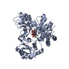


 PDBj
PDBj Assembly
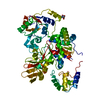

 Type: D-saccharide, beta linking / Mass: 221.208 Da / Num. of mol.: 1
 Mass: 18.015 Da / Num. of mol.: 294 / Source method: isolated from a natural source / Formula: H2O
 Sample preparation
 Processing