 Movie
Movie Controller

[English] 日本語
 Yorodumi
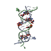
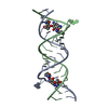
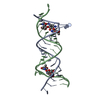
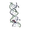
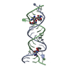
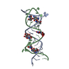
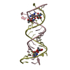
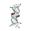
- PDB-5zei: Crystal structure of the bacterial A1408me1A-mutant ribosomal dec... -
+ Open data

- Basic information
| Entry | Database: PDB / ID: 5zei | ||||||||||||||||||||||||
|---|---|---|---|---|---|---|---|---|---|---|---|---|---|---|---|---|---|---|---|---|---|---|---|---|---|
| Title | Crystal structure of the bacterial A1408me1A-mutant ribosomal decoding site in complex with geneticin | ||||||||||||||||||||||||
 Components Components |
| ||||||||||||||||||||||||
 Keywords Keywords | RNA / ribosome / aminoglycoside / antibiotic-resistance | ||||||||||||||||||||||||
| Function / homology | GENETICIN / RNA / RNA (> 10) Function and homology information Function and homology information | ||||||||||||||||||||||||
| Biological species | synthetic construct (others) | ||||||||||||||||||||||||
| Method |  X-RAY DIFFRACTION / SYNCHROTRON / MOLECULAR REPLACEMENT / Resolution: 2.1 Å X-RAY DIFFRACTION / SYNCHROTRON / MOLECULAR REPLACEMENT / Resolution: 2.1 Å | ||||||||||||||||||||||||
 Authors Authors | Kanazawa, H. / Baba, F. / Koganei, M. / Kondo, J. | ||||||||||||||||||||||||
| Funding support |  Japan, 7items Japan, 7items
| ||||||||||||||||||||||||
 Citation Citation | Journal: Nucleic Acids Res. / Year: 2017 Title: A structural basis for the antibiotic resistance conferred by an N1-methylation of A1408 in 16S rRNA. Authors: Kanazawa, H. / Baba, F. / Koganei, M. / Kondo, J. | ||||||||||||||||||||||||
| History |
|
- Structure visualization
| Structure viewer | Molecule: MolmilJmol/JSmol |
|---|
- Downloads & links
-Download
| PDBx/mmCIF format | 5zei.cif.gz | 41.8 KB | Display | PDBx/mmCIF format |
|---|---|---|---|---|
| PDB format | pdb5zei.ent.gz | 27.6 KB | Display | PDB format |
| PDBx/mmJSON format | 5zei.json.gz | Tree view | PDBx/mmJSON format | |
| Others |  Other downloads Other downloads |
-Validation report
| Summary document | 5zei_validation.pdf.gz | 1.1 MB | Display | wwPDB validaton report |
|---|---|---|---|---|
| Full document | 5zei_full_validation.pdf.gz | 1.1 MB | Display | |
| Data in XML | 5zei_validation.xml.gz | 6.2 KB | Display | |
| Data in CIF | 5zei_validation.cif.gz | 8.3 KB | Display | |
| Arichive directory | https://data.pdbj.org/pub/pdb/validation_reports/ze/5zeiftp://data.pdbj.org/pub/pdb/validation_reports/ze/5zei | HTTPS FTP |
-Related structure data
| Related structure data | 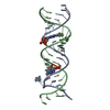 5zegC 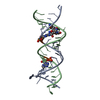 5zejC 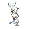 5zemC  3td1S C: citing same article ( S: Starting model for refinement |
|---|---|
| Similar structure data |


-Links
 PDBj
PDBj


- Assembly
| Deposited unit | 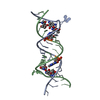
| |||||||||
|---|---|---|---|---|---|---|---|---|---|---|
| 1 |
| |||||||||
| Unit cell |
| |||||||||
| Components on special symmetry positions |
|
-Components
| #1: RNA chain | Mass: 7063.270 Da / Num. of mol.: 1 / Source method: obtained synthetically / Source: (synth.) synthetic construct (others) | ||
|---|---|---|---|
| #2: RNA chain | Mass: 6757.104 Da / Num. of mol.: 1 / Source method: obtained synthetically / Source: (synth.) synthetic construct (others) | ||
| #3: Chemical |   Mass: 496.552 Da / Num. of mol.: 2 / Source method: obtained synthetically / Formula: C20H40N4O10 / Comment: antibiotic*YM Mass: 496.552 Da / Num. of mol.: 2 / Source method: obtained synthetically / Formula: C20H40N4O10 / Comment: antibiotic*YM#4: Water | ChemComp-HOH / |  Mass: 18.015 Da / Num. of mol.: 142 / Source method: isolated from a natural source / Formula: H2O Mass: 18.015 Da / Num. of mol.: 142 / Source method: isolated from a natural source / Formula: H2O |
-Experimental details
-Experiment
| Experiment | Method: X-RAY DIFFRACTION / Number of used crystals: 1 |
|---|
- Sample preparation
| Crystal | Density Matthews: 2.54 Å3/Da / Density % sol: 51.62 % |
|---|---|
| Crystal grow | Temperature: 293 K / Method: vapor diffusion, hanging drop / pH: 7 Details: Sodium cacodylate, Spermine, MPD, ammonium chloride |
-Data collection
| Diffraction | Mean temperature: 100 K |
|---|---|
| Diffraction source | Source: SYNCHROTRON / Site: Photon Factory / Beamline: BL-17A / Wavelength: 0.98 Å |
| Detector | Type: ADSC QUANTUM 315r / Detector: CCD / Date: May 25, 2013 |
| Radiation | Protocol: SINGLE WAVELENGTH / Monochromatic (M) / Laue (L): M / Scattering type: x-ray |
| Radiation wavelength | Wavelength: 0.98 Å / Relative weight: 1 |
| Reflection | Resolution: 2.1→44.9 Å / Num. obs: 8419 / % possible obs: 96.6 % / Redundancy: 6.4 % / Rmerge(I) obs: 0.059 / Net I/σ(I): 15.4 |
| Reflection shell | Resolution: 2.1→2.2 Å / Rmerge(I) obs: 0.365 |
- Processing
| Software |
| |||||||||||||||||||||||||||||||||||||||||||||||||
|---|---|---|---|---|---|---|---|---|---|---|---|---|---|---|---|---|---|---|---|---|---|---|---|---|---|---|---|---|---|---|---|---|---|---|---|---|---|---|---|---|---|---|---|---|---|---|---|---|---|---|
| Refinement | Method to determine structure: MOLECULAR REPLACEMENT Starting model: 3TD1 Resolution: 2.1→44.9 Å / SU ML: 0.26 / Cross valid method: FREE R-VALUE / σ(F): 1.38 / Phase error: 27.39
| |||||||||||||||||||||||||||||||||||||||||||||||||
| Solvent computation | Shrinkage radii: 0.9 Å / VDW probe radii: 1.11 Å | |||||||||||||||||||||||||||||||||||||||||||||||||
| Displacement parameters | Biso max: 92.69 Å2 / Biso mean: 39.21 Å2 / Biso min: 23.86 Å2 | |||||||||||||||||||||||||||||||||||||||||||||||||
| Refinement step | Cycle: final / Resolution: 2.1→44.9 Å
| |||||||||||||||||||||||||||||||||||||||||||||||||
| Refine LS restraints |
| |||||||||||||||||||||||||||||||||||||||||||||||||
| LS refinement shell | Refine-ID: X-RAY DIFFRACTION / Rfactor Rfree error: 0 / Total num. of bins used: 6
|
