+ Open data
- Basic information
| Entry | Database: PDB / ID: 5zap | ||||||
|---|---|---|---|---|---|---|---|
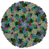
| Title | Atomic structure of the herpes simplex virus type 2 B-capsid | ||||||
 Components Components |
| ||||||
 Keywords Keywords | VIRUS / herpesvirus / capsid / cryo-em / atomic structure | ||||||
| Function / homology |  Function and homology information Function and homology informationT=16 icosahedral viral capsid / viral capsid assembly / viral process / viral capsid / host cell nucleus / structural molecule activity / DNA binding Similarity search - Function | ||||||
| Biological species |   Human herpesvirus 2 Human herpesvirus 2 | ||||||
| Method | ELECTRON MICROSCOPY / single particle reconstruction / cryo EM / Resolution: 3.1 Å | ||||||
 Authors Authors | Yuan, S. / Wang, J.L. / Zhu, D.J. / Wang, N. / Gao, Q. / Chen, W.Y. / Tang, H. / Wang, J.Z. / Zhang, X.Z. / Liu, H.R. ...Yuan, S. / Wang, J.L. / Zhu, D.J. / Wang, N. / Gao, Q. / Chen, W.Y. / Tang, H. / Wang, J.Z. / Zhang, X.Z. / Liu, H.R. / Rao, Z.H. / Wang, X.X. | ||||||
 Citation Citation |  Journal: Science / Year: 2018 Journal: Science / Year: 2018Title: Cryo-EM structure of a herpesvirus capsid at 3.1 Å. Authors: Shuai Yuan / Jialing Wang / Dongjie Zhu / Nan Wang / Qiang Gao / Wenyuan Chen / Hao Tang / Junzhi Wang / Xinzheng Zhang / Hongrong Liu / Zihe Rao / Xiangxi Wang /  Abstract: Structurally and genetically, human herpesviruses are among the largest and most complex of viruses. Using cryo-electron microscopy (cryo-EM) with an optimized image reconstruction strategy, we ...Structurally and genetically, human herpesviruses are among the largest and most complex of viruses. Using cryo-electron microscopy (cryo-EM) with an optimized image reconstruction strategy, we report the herpes simplex virus type 2 (HSV-2) capsid structure at 3.1 angstroms, which is built up of about 3000 proteins organized into three types of hexons (central, peripentonal, and edge), pentons, and triplexes. Both hexons and pentons contain the major capsid protein, VP5; hexons also contain a small capsid protein, VP26; and triplexes comprise VP23 and VP19C. Acting as core organizers, VP5 proteins form extensive intermolecular networks, involving multiple disulfide bonds (about 1500 in total) and noncovalent interactions, with VP26 proteins and triplexes that underpin capsid stability and assembly. Conformational adaptations of these proteins induced by their microenvironments lead to 46 different conformers that assemble into a massive quasisymmetric shell, exemplifying the structural and functional complexity of HSV. #1:  Journal: Nat Commun / Year: 2018 Journal: Nat Commun / Year: 2018Title: Pushing the resolution limit by correcting the Ewald sphere effect in single-particle Cryo-EM reconstructions. Authors: Dongjie Zhu / Xiangxi Wang / Qianglin Fang / James L Van Etten / Michael G Rossmann / Zihe Rao / Xinzheng Zhang /   Abstract: The Ewald sphere effect is generally neglected when using the Central Projection Theorem for cryo electron microscopy single-particle reconstructions. This can reduce the resolution of a ...The Ewald sphere effect is generally neglected when using the Central Projection Theorem for cryo electron microscopy single-particle reconstructions. This can reduce the resolution of a reconstruction. Here we estimate the attainable resolution and report a "block-based" reconstruction method for extending the resolution limit. We find the Ewald sphere effect limits the resolution of large objects, especially large viruses. After processing two real datasets of large viruses, we show that our procedure can extend the resolution for both datasets and can accommodate the flexibility associated with large protein complexes. | ||||||
| History |
|
- Structure visualization
| Movie |
 Movie viewer Movie viewer |
|---|---|
| Structure viewer | Molecule:  Molmil Molmil Jmol/JSmol Jmol/JSmol |
- Downloads & links
- Download
| PDBx/mmCIF format |  5zap.cif.gz 5zap.cif.gz | 4.4 MB | Display |  PDBx/mmCIF format PDBx/mmCIF format |
|---|---|---|---|---|
| PDB format |  pdb5zap.ent.gz pdb5zap.ent.gz | Display |  PDB format PDB format | |
| PDBx/mmJSON format |  5zap.json.gz 5zap.json.gz | Tree view |  PDBx/mmJSON format PDBx/mmJSON format | |
| Others |  Other downloads Other downloads |
-Validation report
| Summary document |  5zap_validation.pdf.gz 5zap_validation.pdf.gz | 1.4 MB | Display |  wwPDB validaton report wwPDB validaton report |
|---|---|---|---|---|
| Full document |  5zap_full_validation.pdf.gz 5zap_full_validation.pdf.gz | 1.6 MB | Display | |
| Data in XML |  5zap_validation.xml.gz 5zap_validation.xml.gz | 613.2 KB | Display | |
| Data in CIF |  5zap_validation.cif.gz 5zap_validation.cif.gz | 989.9 KB | Display | |
| Arichive directory |  https://data.pdbj.org/pub/pdb/validation_reports/za/5zap https://data.pdbj.org/pub/pdb/validation_reports/za/5zap ftp://data.pdbj.org/pub/pdb/validation_reports/za/5zap ftp://data.pdbj.org/pub/pdb/validation_reports/za/5zap | HTTPS FTP |
-Related structure data
| Related structure data |  6907MC M: map data used to model this data C: citing same article ( |
|---|---|
| Similar structure data |
- Links
Links
- Assembly
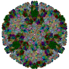
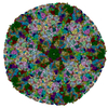
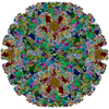
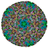
| Deposited unit | 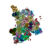
|
|---|---|
| 1 | x 60
|
| 2 |
|
| 3 | x 5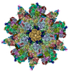
|
| 4 | x 6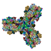
|
| 5 | 
|
| Symmetry | Point symmetry: (Schoenflies symbol: I (icosahedral)) |
- Components
| #1: Protein | Mass: 149370.359 Da / Num. of mol.: 16 / Source method: isolated from a natural source / Source: (natural)   Human herpesvirus 2 / References: UniProt: G9I240 Human herpesvirus 2 / References: UniProt: G9I240#2: Protein | Mass: 34373.785 Da / Num. of mol.: 10 / Source method: isolated from a natural source / Source: (natural)   Human herpesvirus 2 / References: UniProt: G9I239 Human herpesvirus 2 / References: UniProt: G9I239#3: Protein | Mass: 50542.617 Da / Num. of mol.: 5 / Source method: isolated from a natural source / Source: (natural)   Human herpesvirus 2 / References: UniProt: A0A1U9ZFW1 Human herpesvirus 2 / References: UniProt: A0A1U9ZFW1#4: Protein | Mass: 12147.707 Da / Num. of mol.: 15 / Source method: isolated from a natural source / Source: (natural)   Human herpesvirus 2 / References: UniProt: G9I257 Human herpesvirus 2 / References: UniProt: G9I257Has protein modification | Y | |
|---|
-Experimental details
-Experiment
| Experiment | Method: ELECTRON MICROSCOPY |
|---|---|
| EM experiment | Aggregation state: PARTICLE / 3D reconstruction method: single particle reconstruction |
- Sample preparation
| Component | Name: Human herpesvirus 2 / Type: VIRUS / Entity ID: #1, #4, #2-#3 / Source: NATURAL |
|---|---|
| Source (natural) | Organism:   Human herpesvirus 2 Human herpesvirus 2 |
| Details of virus | Empty: NO / Enveloped: NO / Isolate: STRAIN / Type: VIRION |
| Natural host | Organism: Human herpesvirus 2 |
| Buffer solution | pH: 7.4 |
| Specimen | Embedding applied: NO / Shadowing applied: NO / Staining applied: NO / Vitrification applied: YES |
| Vitrification | Cryogen name: ETHANE |
- Electron microscopy imaging
| Experimental equipment |  Model: Titan Krios / Image courtesy: FEI Company |
|---|---|
| Microscopy | Model: FEI TITAN KRIOS |
| Electron gun | Electron source:  FIELD EMISSION GUN / Accelerating voltage: 300 kV / Illumination mode: FLOOD BEAM FIELD EMISSION GUN / Accelerating voltage: 300 kV / Illumination mode: FLOOD BEAM |
| Electron lens | Mode: BRIGHT FIELD |
| Image recording | Electron dose: 1.1 e/Å2 / Film or detector model: FEI FALCON II (4k x 4k) |
- Processing
| CTF correction | Type: NONE |
|---|---|
| 3D reconstruction | Resolution: 3.1 Å / Resolution method: FSC 0.143 CUT-OFF / Num. of particles: 45000 / Symmetry type: POINT |
 Movie
Movie Controller





 PDBj
PDBj
