 Movie
Movie Controller

[English] 日本語
 Yorodumi
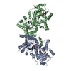
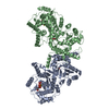
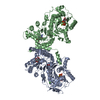
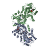
- PDB-5z79: Crystal Structure Analysis of the HPPK-DHPS in complex with substrates -
+ Open data

- Basic information
| Entry | Database: PDB / ID: 5z79 | ||||||
|---|---|---|---|---|---|---|---|
| Title | Crystal Structure Analysis of the HPPK-DHPS in complex with substrates | ||||||
 Components Components | Hydroxymethyldihydropterin pyrophosphokinase-dihydropteroate synthase, putative | ||||||
 Keywords Keywords | TRANSFERASE / Bifunctional / Malaria Pterin / Substrates | ||||||
| Function / homology |  Function and homology information Function and homology information2-amino-4-hydroxy-6-hydroxymethyldihydropteridine diphosphokinase / 2-amino-4-hydroxy-6-hydroxymethyldihydropteridine diphosphokinase activity / dihydropteroate synthase activity / folic acid biosynthetic process / tetrahydrofolate biosynthetic process / kinase activity / ATP binding / metal ion binding Similarity search - Function | ||||||
| Biological species |  | ||||||
| Method |  X-RAY DIFFRACTION / SYNCHROTRON / MOLECULAR REPLACEMENT / molecular replacement / Resolution: 2.9 Å X-RAY DIFFRACTION / SYNCHROTRON / MOLECULAR REPLACEMENT / molecular replacement / Resolution: 2.9 Å | ||||||
 Authors Authors | Manickam, Y. / Karl, H. / Sharma, A. | ||||||
 Citation Citation | Journal: J. Biol. Chem. / Year: 2018 Title: Structure of 6-hydroxymethyl-7,8-dihydropterin pyrophosphokinase-dihydropteroate synthase fromPlasmodium vivaxsheds light on drug resistance Authors: Yogavel, M. / Nettleship, J.E. / Sharma, A. / Harlos, K. / Jamwal, A. / Chaturvedi, R. / Sharma, M. / Jain, V. / Chhibber-Goel, J. / Sharma, A. | ||||||
| History |
|
- Structure visualization
| Structure viewer | Molecule: MolmilJmol/JSmol |
|---|
- Downloads & links
-Download
| PDBx/mmCIF format | 5z79.cif.gz | 1.4 MB | Display | PDBx/mmCIF format |
|---|---|---|---|---|
| PDB format | pdb5z79.ent.gz | 1.1 MB | Display | PDB format |
| PDBx/mmJSON format | 5z79.json.gz | Tree view | PDBx/mmJSON format | |
| Others |  Other downloads Other downloads |
-Validation report
| Arichive directory | https://data.pdbj.org/pub/pdb/validation_reports/z7/5z79ftp://data.pdbj.org/pub/pdb/validation_reports/z7/5z79 | HTTPS FTP |
|---|
-Related structure data
| Related structure data |  1eyeS S: Starting model for refinement |
|---|---|
| Similar structure data |

-Links
 PDBj
PDBj


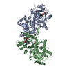
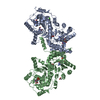
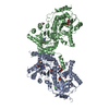
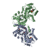
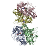
- Assembly
| Deposited unit | 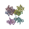
| ||||||||
|---|---|---|---|---|---|---|---|---|---|
| 1 | 
| ||||||||
| 2 | 
| ||||||||
| 3 | 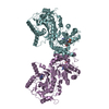
| ||||||||
| Unit cell |
|
-Components
-Protein , 1 types, 6 molecules ABCDEF
| #1: Protein | Mass: 82484.242 Da / Num. of mol.: 6 / Source method: obtained synthetically Source: (synth.) References: UniProt: A0A1K9YMY7, UniProt: A5JZS1*PLUS, dihydropteroate synthase, 2-amino-4-hydroxy-6-hydroxymethyldihydropteridine diphosphokinase |
|---|
-Non-polymers , 7 types, 241 molecules 


| #2: Chemical | ChemComp-HH2 /  Mass: 353.123 Da / Num. of mol.: 6 / Source method: obtained synthetically / Formula: C7H9N5O8P2 Mass: 353.123 Da / Num. of mol.: 6 / Source method: obtained synthetically / Formula: C7H9N5O8P2#3: Chemical |  Mass: 505.208 Da / Num. of mol.: 2 / Source method: obtained synthetically / Formula: C11H18N5O12P3 / Comment: AMP-CPP, energy-carrying molecule analogue*YM Mass: 505.208 Da / Num. of mol.: 2 / Source method: obtained synthetically / Formula: C11H18N5O12P3 / Comment: AMP-CPP, energy-carrying molecule analogue*YM#4: Chemical | ChemComp-PAB /  Mass: 137.136 Da / Num. of mol.: 6 / Source method: obtained synthetically / Formula: C7H7NO2 Mass: 137.136 Da / Num. of mol.: 6 / Source method: obtained synthetically / Formula: C7H7NO2#5: Chemical | ChemComp-MG /  Mass: 24.305 Da / Num. of mol.: 4 / Source method: obtained synthetically / Formula: Mg Mass: 24.305 Da / Num. of mol.: 4 / Source method: obtained synthetically / Formula: Mg#6: Chemical | ChemComp-PE0 /  Mass: 163.137 Da / Num. of mol.: 4 / Source method: obtained synthetically / Formula: C6H5N5O Mass: 163.137 Da / Num. of mol.: 4 / Source method: obtained synthetically / Formula: C6H5N5O#7: Chemical |  Mass: 251.242 Da / Num. of mol.: 2 / Source method: obtained synthetically / Formula: C10H13N5O3 Mass: 251.242 Da / Num. of mol.: 2 / Source method: obtained synthetically / Formula: C10H13N5O3#8: Water | ChemComp-HOH / | Mass: 18.015 Da / Num. of mol.: 217 / Source method: isolated from a natural source / Formula: H2O |
|---|
-Details
| Has protein modification | N |
|---|---|
| Sequence details | The used cloned protein sequence has Ala383. |
-Experimental details
-Experiment
| Experiment | Method: X-RAY DIFFRACTION / Number of used crystals: 1 |
|---|
- Sample preparation
| Crystal | Density Matthews: 2.69 Å3/Da / Density % sol: 54.3 % |
|---|---|
| Crystal grow | Temperature: 293 K / Method: vapor diffusion, hanging drop Details: 20% PEG3350, 0.2M Potassium citrate tribasic monohydrate |
-Data collection
| Diffraction | Mean temperature: 100 K |
|---|---|
| Diffraction source | Source: SYNCHROTRON / Site: Diamond  / Beamline: I03 / Wavelength: 0.9763 Å / Beamline: I03 / Wavelength: 0.9763 Å |
| Detector | Type: DECTRIS PILATUS3 S 6M / Detector: PIXEL / Date: Oct 10, 2017 |
| Radiation | Monochromator: SINGLE WAVELENGTH / Protocol: SINGLE WAVELENGTH / Monochromatic (M) / Laue (L): M / Scattering type: x-ray |
| Radiation wavelength | Wavelength: 0.9763 Å / Relative weight: 1 |
| Reflection | Resolution: 2.79→49.82 Å / Num. obs: 129475 / % possible obs: 99.4 % / Redundancy: 3.5 % / CC1/2: 0.9 / Net I/σ(I): 4.5 |
| Reflection shell | Resolution: 2.79→2.84 Å / Mean I/σ(I) obs: 0.7 / Num. unique obs: 5799 / CC1/2: 0.2 / % possible all: 90.41 |
-Phasing
| Phasing | Method: molecular replacement |
|---|
- Processing
| Software |
| ||||||||||||||||||||||||||||||||||||||||||||||||||||||||||||||||||||||||||||||||||||||||||||||||||||||||||||||||||||||||||||||||||||||||||||||||||||||||||||||||||||||||||||||||||||||
|---|---|---|---|---|---|---|---|---|---|---|---|---|---|---|---|---|---|---|---|---|---|---|---|---|---|---|---|---|---|---|---|---|---|---|---|---|---|---|---|---|---|---|---|---|---|---|---|---|---|---|---|---|---|---|---|---|---|---|---|---|---|---|---|---|---|---|---|---|---|---|---|---|---|---|---|---|---|---|---|---|---|---|---|---|---|---|---|---|---|---|---|---|---|---|---|---|---|---|---|---|---|---|---|---|---|---|---|---|---|---|---|---|---|---|---|---|---|---|---|---|---|---|---|---|---|---|---|---|---|---|---|---|---|---|---|---|---|---|---|---|---|---|---|---|---|---|---|---|---|---|---|---|---|---|---|---|---|---|---|---|---|---|---|---|---|---|---|---|---|---|---|---|---|---|---|---|---|---|---|---|---|---|---|
| Refinement | Method to determine structure: MOLECULAR REPLACEMENT Starting model: 1EYE Resolution: 2.9→49.82 Å / Cor.coef. Fo:Fc: 0.89 / Cor.coef. Fo:Fc free: 0.84 / SU B: 41.744 / SU ML: 0.371 / Cross valid method: THROUGHOUT / ESU R Free: 0.423 / Details: HYDROGENS HAVE BEEN ADDED IN THE RIDING POSITIONS
| ||||||||||||||||||||||||||||||||||||||||||||||||||||||||||||||||||||||||||||||||||||||||||||||||||||||||||||||||||||||||||||||||||||||||||||||||||||||||||||||||||||||||||||||||||||||
| Solvent computation | Ion probe radii: 0.8 Å / Shrinkage radii: 0.8 Å / VDW probe radii: 1.2 Å | ||||||||||||||||||||||||||||||||||||||||||||||||||||||||||||||||||||||||||||||||||||||||||||||||||||||||||||||||||||||||||||||||||||||||||||||||||||||||||||||||||||||||||||||||||||||
| Displacement parameters | Biso mean: 43.362 Å2
| ||||||||||||||||||||||||||||||||||||||||||||||||||||||||||||||||||||||||||||||||||||||||||||||||||||||||||||||||||||||||||||||||||||||||||||||||||||||||||||||||||||||||||||||||||||||
| Refinement step | Cycle: 1 / Resolution: 2.9→49.82 Å
| ||||||||||||||||||||||||||||||||||||||||||||||||||||||||||||||||||||||||||||||||||||||||||||||||||||||||||||||||||||||||||||||||||||||||||||||||||||||||||||||||||||||||||||||||||||||
| Refine LS restraints |
|