 Movie
Movie Controller


+ Open data

- Basic information
| Entry | Database: PDB / ID: 6jwq | ||||||
|---|---|---|---|---|---|---|---|
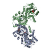
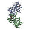
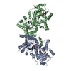
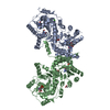
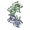
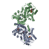
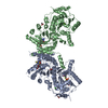
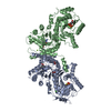
| Title | Crystal structure of Plasmodium falciparum HPPK-DHPS wild type | ||||||
 Components Components | 7,8-dihydro-6-hydroxymethylpterin pyrophosphokinase-dihydropteroate synthase | ||||||
 Keywords Keywords | TRANSFERASE / TIM barrel / kinase | ||||||
| Function / homology |  Function and homology information Function and homology information2-amino-4-hydroxy-6-hydroxymethyldihydropteridine diphosphokinase activity / dihydropteroate synthase activity / mitochondrial envelope / folic acid biosynthetic process / tetrahydrofolate biosynthetic process / kinase activity / ATP binding / metal ion binding Similarity search - Function | ||||||
| Biological species |  | ||||||
| Method |  X-RAY DIFFRACTION / SYNCHROTRON / SAD / Resolution: 2.5 Å X-RAY DIFFRACTION / SYNCHROTRON / SAD / Resolution: 2.5 Å | ||||||
| Model details | Crystal structure of Plasmodium falciparum HPPK-DHPS apo form | ||||||
 Authors Authors | Chitnumsub, P. / Jaruwat, A. / Yuthavong, Y. | ||||||
| Funding support |  Thailand, 1items Thailand, 1items
| ||||||
 Citation Citation | Journal: Febs J. / Year: 2020 Title: The structure of Plasmodium falciparum hydroxymethyldihydropterin pyrophosphokinase-dihydropteroate synthase reveals the basis of sulfa resistance. Authors: Chitnumsub, P. / Jaruwat, A. / Talawanich, Y. / Noytanom, K. / Liwnaree, B. / Poen, S. / Yuthavong, Y. | ||||||
| History |
|
- Structure visualization
| Structure viewer | Molecule: MolmilJmol/JSmol |
|---|
- Downloads & links
-Download
| PDBx/mmCIF format | 6jwq.cif.gz | 251.5 KB | Display | PDBx/mmCIF format |
|---|---|---|---|---|
| PDB format | pdb6jwq.ent.gz | 197.9 KB | Display | PDB format |
| PDBx/mmJSON format | 6jwq.json.gz | Tree view | PDBx/mmJSON format | |
| Others |  Other downloads Other downloads |
-Validation report
| Arichive directory | https://data.pdbj.org/pub/pdb/validation_reports/jw/6jwqftp://data.pdbj.org/pub/pdb/validation_reports/jw/6jwq | HTTPS FTP |
|---|
-Related structure data
| Related structure data |  6jwrC  6jwsC  6jwtC  6jwuC  6jwvC  6jwwC  6jwxC  6jwyC  6jwzC  6kckC 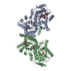 6kclC  6kcmC C: citing same article ( |
|---|---|
| Similar structure data |


-Links
 PDBj
PDBj


- Assembly
| Deposited unit | 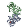
| ||||||||
|---|---|---|---|---|---|---|---|---|---|
| 1 |
| ||||||||
| Unit cell |
|
-Components
-Protein , 1 types, 2 molecules AB
| #1: Protein | Mass: 85957.492 Da / Num. of mol.: 2 / Fragment: HPPK Source method: isolated from a genetically manipulated source Source: (gene. exp.) Gene: PPPK-DHPS / Plasmid: pET29a / Production host:  |
|---|
-Non-polymers , 6 types, 137 molecules 


| #2: Chemical |  Mass: 24.305 Da / Num. of mol.: 2 / Fragment: DHPS / Source method: obtained synthetically / Formula: Mg / References: dihydropteroate synthase Mass: 24.305 Da / Num. of mol.: 2 / Fragment: DHPS / Source method: obtained synthetically / Formula: Mg / References: dihydropteroate synthase#3: Chemical | ChemComp-ATP / |  Mass: 507.181 Da / Num. of mol.: 1 / Source method: obtained synthetically / Formula: C10H16N5O13P3 / Comment: ATP, energy-carrying molecule*YM Mass: 507.181 Da / Num. of mol.: 1 / Source method: obtained synthetically / Formula: C10H16N5O13P3 / Comment: ATP, energy-carrying molecule*YM#4: Chemical | ChemComp-CA / |  Mass: 40.078 Da / Num. of mol.: 1 / Source method: obtained synthetically / Formula: Ca Mass: 40.078 Da / Num. of mol.: 1 / Source method: obtained synthetically / Formula: Ca#5: Chemical |  Mass: 59.044 Da / Num. of mol.: 2 / Source method: obtained synthetically / Formula: C2H3O2 Mass: 59.044 Da / Num. of mol.: 2 / Source method: obtained synthetically / Formula: C2H3O2#6: Chemical | ChemComp-AMP / |  Mass: 347.221 Da / Num. of mol.: 1 / Source method: obtained synthetically / Formula: C10H14N5O7P / Comment: AMP*YM Mass: 347.221 Da / Num. of mol.: 1 / Source method: obtained synthetically / Formula: C10H14N5O7P / Comment: AMP*YM#7: Water | ChemComp-HOH / | Mass: 18.015 Da / Num. of mol.: 130 / Source method: isolated from a natural source / Formula: H2O |
|---|
-Experimental details
-Experiment
| Experiment | Method: X-RAY DIFFRACTION / Number of used crystals: 1 |
|---|
- Sample preparation
| Crystal | Density Matthews: 2.81 Å3/Da / Density % sol: 56.26 % / Mosaicity: 0.751 ° |
|---|---|
| Crystal grow | Temperature: 298 K / Method: microbatch / pH: 9 Details: 0.1 M bicine buffer pH 9.0, 0.5-0.6 M Ca acetate and 20%w/v PEG4000 |
-Data collection
| Diffraction | Mean temperature: 100 K / Serial crystal experiment: N | |||||||||||||||||||||||||||||||||||||||||||||||||||||||||||||||||||||||||||||
|---|---|---|---|---|---|---|---|---|---|---|---|---|---|---|---|---|---|---|---|---|---|---|---|---|---|---|---|---|---|---|---|---|---|---|---|---|---|---|---|---|---|---|---|---|---|---|---|---|---|---|---|---|---|---|---|---|---|---|---|---|---|---|---|---|---|---|---|---|---|---|---|---|---|---|---|---|---|---|
| Diffraction source | Source: SYNCHROTRON / Site: NSRRC  / Beamline: BL13B1 / Wavelength: 1 Å / Beamline: BL13B1 / Wavelength: 1 Å | |||||||||||||||||||||||||||||||||||||||||||||||||||||||||||||||||||||||||||||
| Detector | Type: ADSC QUANTUM 315 / Detector: CCD / Date: Mar 11, 2010 | |||||||||||||||||||||||||||||||||||||||||||||||||||||||||||||||||||||||||||||
| Radiation | Protocol: SINGLE WAVELENGTH / Monochromatic (M) / Laue (L): M / Scattering type: x-ray | |||||||||||||||||||||||||||||||||||||||||||||||||||||||||||||||||||||||||||||
| Radiation wavelength | Wavelength: 1 Å / Relative weight: 1 | |||||||||||||||||||||||||||||||||||||||||||||||||||||||||||||||||||||||||||||
| Reflection | Resolution: 2.5→30 Å / Num. obs: 65642 / % possible obs: 97.3 % / Redundancy: 4.7 % / Rmerge(I) obs: 0.056 / Χ2: 1.065 / Net I/σ(I): 15.3 / Num. measured all: 307040 | |||||||||||||||||||||||||||||||||||||||||||||||||||||||||||||||||||||||||||||
| Reflection shell |
|
- Processing
| Software |
| |||||||||||||||||||||||||||||||||||||||||||||
|---|---|---|---|---|---|---|---|---|---|---|---|---|---|---|---|---|---|---|---|---|---|---|---|---|---|---|---|---|---|---|---|---|---|---|---|---|---|---|---|---|---|---|---|---|---|---|
| Refinement | Method to determine structure: SAD / Resolution: 2.5→30 Å / Cor.coef. Fo:Fc: 0.945 / Cor.coef. Fo:Fc free: 0.917 / SU B: 9.819 / SU ML: 0.212 / Cross valid method: THROUGHOUT / σ(F): 0 / ESU R: 0.368 / ESU R Free: 0.272 Details: HYDROGENS HAVE BEEN USED IF PRESENT IN THE INPUT U VALUES : REFINED INDIVIDUALLY
| |||||||||||||||||||||||||||||||||||||||||||||
| Solvent computation | Ion probe radii: 0.8 Å / Shrinkage radii: 0.8 Å / VDW probe radii: 1.4 Å | |||||||||||||||||||||||||||||||||||||||||||||
| Displacement parameters | Biso max: 120 Å2 / Biso mean: 63.656 Å2 / Biso min: 21.17 Å2
| |||||||||||||||||||||||||||||||||||||||||||||
| Refinement step | Cycle: final / Resolution: 2.5→30 Å
| |||||||||||||||||||||||||||||||||||||||||||||
| Refine LS restraints |
| |||||||||||||||||||||||||||||||||||||||||||||
| LS refinement shell | Resolution: 2.503→2.567 Å / Rfactor Rfree error: 0 / Total num. of bins used: 20
|
