 Movie
Movie Controller


+ Open data

- Basic information
| Entry | Database: PDB / ID: 5z28 | ||||||
|---|---|---|---|---|---|---|---|
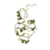
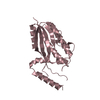
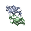
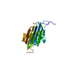
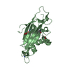
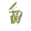
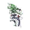
| Title | AtVAL2 PHD-Like domain | ||||||
 Components Components | B3 domain-containing transcription repressor VAL2 | ||||||
 Keywords Keywords | TRANSCRIPTION / transcriptional factor / Val2 / PHD-Like domain / FLC / plant | ||||||
| Function / homology |  Function and homology information Function and homology informationtranscription cis-regulatory region binding / DNA-binding transcription factor activity / regulation of DNA-templated transcription / zinc ion binding / nucleus Similarity search - Function | ||||||
| Biological species |  | ||||||
| Method |  X-RAY DIFFRACTION / SYNCHROTRON / MOLECULAR REPLACEMENT / Resolution: 2.245 Å X-RAY DIFFRACTION / SYNCHROTRON / MOLECULAR REPLACEMENT / Resolution: 2.245 Å | ||||||
 Authors Authors | Wu, B.X. / Zhang, M.M. | ||||||
 Citation Citation | Journal: To Be Published Title: Crystal structure of AtVAL12 PHD-Like domain Authors: Wu, B.X. / Zhang, M.M. | ||||||
| History |
|
- Structure visualization
| Structure viewer | Molecule: MolmilJmol/JSmol |
|---|
- Downloads & links
-Download
| PDBx/mmCIF format | 5z28.cif.gz | 51.6 KB | Display | PDBx/mmCIF format |
|---|---|---|---|---|
| PDB format | pdb5z28.ent.gz | 35 KB | Display | PDB format |
| PDBx/mmJSON format | 5z28.json.gz | Tree view | PDBx/mmJSON format | |
| Others |  Other downloads Other downloads |
-Validation report
| Arichive directory | https://data.pdbj.org/pub/pdb/validation_reports/z2/5z28ftp://data.pdbj.org/pub/pdb/validation_reports/z2/5z28 | HTTPS FTP |
|---|
-Related structure data
| Related structure data |  5yugS S: Starting model for refinement |
|---|---|
| Similar structure data |


-Links
 PDBj
PDBj- Assembly
| Deposited unit | 
| ||||||||
|---|---|---|---|---|---|---|---|---|---|
| 1 |
| ||||||||
| Unit cell |
|
-Components
| #1: Protein | Mass: 11156.031 Da / Num. of mol.: 2 / Fragment: PHD-Like domain Source method: isolated from a genetically manipulated source Source: (gene. exp.)  #2: Chemical | ChemComp-ZN /   Mass: 65.409 Da / Num. of mol.: 6 / Source method: obtained synthetically / Formula: Zn Mass: 65.409 Da / Num. of mol.: 6 / Source method: obtained synthetically / Formula: Zn#3: Chemical | ChemComp-GOL / |   Mass: 92.094 Da / Num. of mol.: 1 / Source method: obtained synthetically / Formula: C3H8O3 Mass: 92.094 Da / Num. of mol.: 1 / Source method: obtained synthetically / Formula: C3H8O3#4: Water | ChemComp-HOH / |  Mass: 18.015 Da / Num. of mol.: 22 / Source method: isolated from a natural source / Formula: H2O Mass: 18.015 Da / Num. of mol.: 22 / Source method: isolated from a natural source / Formula: H2O |
|---|
-Experimental details
-Experiment
| Experiment | Method: X-RAY DIFFRACTION / Number of used crystals: 1 |
|---|
- Sample preparation
| Crystal | Density Matthews: 2.32 Å3/Da / Density % sol: 46.93 % |
|---|---|
| Crystal grow | Temperature: 293 K / Method: vapor diffusion, sitting drop / Details: 0.1M Bis-Tris pH6.5, 28% PEG MME 2000 |
-Data collection
| Diffraction | Mean temperature: 100 K |
|---|---|
| Diffraction source | Source: SYNCHROTRON / Site: SSRF  / Beamline: BL18U1 / Wavelength: 0.97853 Å / Beamline: BL18U1 / Wavelength: 0.97853 Å |
| Detector | Type: DECTRIS PILATUS3 S 6M / Detector: PIXEL / Date: Dec 29, 2017 |
| Radiation | Protocol: SINGLE WAVELENGTH / Monochromatic (M) / Laue (L): M / Scattering type: x-ray |
| Radiation wavelength | Wavelength: 0.97853 Å / Relative weight: 1 |
| Reflection | Resolution: 2.244→52.11 Å / Num. obs: 10184 / % possible obs: 99.6 % / Redundancy: 9.6 % / Rmerge(I) obs: 0.147 / Net I/σ(I): 17.94 |
| Reflection shell | Resolution: 2.25→2.33 Å / Redundancy: 6.6 % |
- Processing
| Software |
| |||||||||||||||||||||||||||||||||||
|---|---|---|---|---|---|---|---|---|---|---|---|---|---|---|---|---|---|---|---|---|---|---|---|---|---|---|---|---|---|---|---|---|---|---|---|---|
| Refinement | Method to determine structure: MOLECULAR REPLACEMENT Starting model: 5YUG Resolution: 2.245→26.882 Å / SU ML: 0.25 / Cross valid method: FREE R-VALUE / σ(F): 1.39 / Phase error: 23.75
| |||||||||||||||||||||||||||||||||||
| Solvent computation | Shrinkage radii: 0.9 Å / VDW probe radii: 1.11 Å | |||||||||||||||||||||||||||||||||||
| Refinement step | Cycle: LAST / Resolution: 2.245→26.882 Å
| |||||||||||||||||||||||||||||||||||
| Refine LS restraints |
| |||||||||||||||||||||||||||||||||||
| LS refinement shell |
|
