 Movie
Movie Controller

[English] 日本語
 Yorodumi
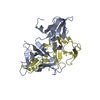
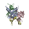
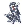
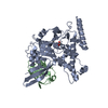
- PDB-5yuo: Crystal structure of SSB protein from Pseudomonas aeruginosa PAO1 -
+ Open data

- Basic information
| Entry | Database: PDB / ID: 5yuo | ||||||
|---|---|---|---|---|---|---|---|
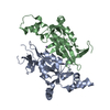
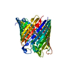
| Title | Crystal structure of SSB protein from Pseudomonas aeruginosa PAO1 | ||||||
 Components Components | Single-stranded DNA-binding protein | ||||||
 Keywords Keywords | DNA BINDING PROTEIN / single-strand DNA binding protein / Pseudomonas aeruginosa | ||||||
| Function / homology |  Function and homology information Function and homology informationnucleoid / enzyme activator activity / single-stranded DNA binding / DNA recombination / DNA replication / DNA repair Similarity search - Function | ||||||
| Biological species |  Pseudomonas aeruginosa PAO1 (bacteria) Pseudomonas aeruginosa PAO1 (bacteria) | ||||||
| Method |  X-RAY DIFFRACTION / SYNCHROTRON / MOLECULAR REPLACEMENT / Resolution: 2.037 Å X-RAY DIFFRACTION / SYNCHROTRON / MOLECULAR REPLACEMENT / Resolution: 2.037 Å | ||||||
 Authors Authors | Huang, Y.H. / Huang, C.Y. | ||||||
 Citation Citation | Journal: RSC Adv. / Year: 2018 Title: The glycine-rich flexible region in SSB is crucial for PriA stimulation. Authors: Huang, Y.H. / Huang, C.Y. | ||||||
| History |
|
- Structure visualization
| Structure viewer | Molecule: MolmilJmol/JSmol |
|---|
- Downloads & links
-Download
| PDBx/mmCIF format | 5yuo.cif.gz | 95.4 KB | Display | PDBx/mmCIF format |
|---|---|---|---|---|
| PDB format | pdb5yuo.ent.gz | 72.7 KB | Display | PDB format |
| PDBx/mmJSON format | 5yuo.json.gz | Tree view | PDBx/mmJSON format | |
| Others |  Other downloads Other downloads |
-Validation report
| Arichive directory | https://data.pdbj.org/pub/pdb/validation_reports/yu/5yuoftp://data.pdbj.org/pub/pdb/validation_reports/yu/5yuo | HTTPS FTP |
|---|
-Related structure data
| Related structure data |  1eygS S: Starting model for refinement |
|---|---|
| Similar structure data |


-Links
 PDBj
PDBj- Assembly
| Deposited unit | 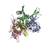
| ||||||||
|---|---|---|---|---|---|---|---|---|---|
| 1 |
| ||||||||
| Unit cell |
| ||||||||
| Components on special symmetry positions |
|
-Components
| #1: Protein | Mass: 13774.341 Da / Num. of mol.: 4 Source method: isolated from a genetically manipulated source Source: (gene. exp.) Pseudomonas aeruginosa PAO1 (bacteria)Strain: ATCC 15692 / DSM 22644 / CIP 104116 / JCM 14847 / LMG 12228 / 1C / PRS 101 / PAO1 Gene: ssb, PA4232 / Production host: #2: Chemical |   Mass: 92.094 Da / Num. of mol.: 2 / Source method: isolated from a natural source / Formula: C3H8O3 Mass: 92.094 Da / Num. of mol.: 2 / Source method: isolated from a natural source / Formula: C3H8O3#3: Water | ChemComp-HOH / |  Mass: 18.015 Da / Num. of mol.: 87 / Source method: isolated from a natural source / Formula: H2O Mass: 18.015 Da / Num. of mol.: 87 / Source method: isolated from a natural source / Formula: H2O |
|---|
-Experimental details
-Experiment
| Experiment | Method: X-RAY DIFFRACTION / Number of used crystals: 1 |
|---|
- Sample preparation
| Crystal | Density Matthews: 2.66 Å3/Da / Density % sol: 53.73 % |
|---|---|
| Crystal grow | Temperature: 298 K / Method: vapor diffusion, hanging drop / pH: 7 Details: 14% PEG 4000, 100mM HEPES pH 7.0, 50mM sodium acetate |
-Data collection
| Diffraction | Mean temperature: 298 K |
|---|---|
| Diffraction source | Source: SYNCHROTRON / Site: NSRRC  / Beamline: BL13C1 / Wavelength: 0.975 Å / Beamline: BL13C1 / Wavelength: 0.975 Å |
| Detector | Type: ADSC QUANTUM 210 / Detector: CCD / Date: Mar 2, 2016 |
| Radiation | Protocol: SINGLE WAVELENGTH / Monochromatic (M) / Laue (L): M / Scattering type: x-ray |
| Radiation wavelength | Wavelength: 0.975 Å / Relative weight: 1 |
| Reflection | Resolution: 2.037→30 Å / Num. obs: 36409 / % possible obs: 98.2 % / Redundancy: 3.6 % / Rmerge(I) obs: 0.038 / Net I/σ(I): 31.36 |
| Reflection shell | Resolution: 2.04→2.11 Å / Rmerge(I) obs: 0.456 / Mean I/σ(I) obs: 2.53 / Num. unique obs: 3312 / % possible all: 89.9 |
- Processing
| Software |
| ||||||||||||||||||||||||||||||||||||||||||||||||||||||||||||||||||||||||||||||||||||||||||||||||||
|---|---|---|---|---|---|---|---|---|---|---|---|---|---|---|---|---|---|---|---|---|---|---|---|---|---|---|---|---|---|---|---|---|---|---|---|---|---|---|---|---|---|---|---|---|---|---|---|---|---|---|---|---|---|---|---|---|---|---|---|---|---|---|---|---|---|---|---|---|---|---|---|---|---|---|---|---|---|---|---|---|---|---|---|---|---|---|---|---|---|---|---|---|---|---|---|---|---|---|---|
| Refinement | Method to determine structure: MOLECULAR REPLACEMENT Starting model: 1EYG Resolution: 2.037→26.49 Å / SU ML: 0.26 / Cross valid method: THROUGHOUT / σ(F): 1.35 / Phase error: 30.44
| ||||||||||||||||||||||||||||||||||||||||||||||||||||||||||||||||||||||||||||||||||||||||||||||||||
| Solvent computation | Shrinkage radii: 0.9 Å / VDW probe radii: 1.11 Å | ||||||||||||||||||||||||||||||||||||||||||||||||||||||||||||||||||||||||||||||||||||||||||||||||||
| Refinement step | Cycle: LAST / Resolution: 2.037→26.49 Å
| ||||||||||||||||||||||||||||||||||||||||||||||||||||||||||||||||||||||||||||||||||||||||||||||||||
| Refine LS restraints |
| ||||||||||||||||||||||||||||||||||||||||||||||||||||||||||||||||||||||||||||||||||||||||||||||||||
| LS refinement shell |
|
