 Movie
Movie Controller


+ Open data

- Basic information
| Entry | Database: PDB / ID: 5ysx | ||||||
|---|---|---|---|---|---|---|---|
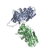
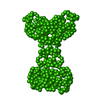
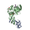
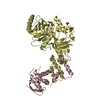
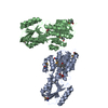
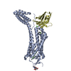
| Title | Structure of P domain of GII.2 Noroviruses | ||||||
 Components Components | VP1 | ||||||
 Keywords Keywords | CELL ADHESION / GII.2 Noroviruses / P dimer | ||||||
| Function / homology |  Function and homology information Function and homology informationNucleoplasmin-like/VP (viral coat and capsid proteins) / Positive stranded ssRNA viruses / Positive stranded ssRNA viruses / Calicivirus coat protein C-terminal / Calicivirus coat protein C-terminal / Calicivirus coat protein / Calicivirus coat protein / Elongation Factor Tu (Ef-tu); domain 3 / Picornavirus/Calicivirus coat protein / Viral coat protein subunit ...Nucleoplasmin-like/VP (viral coat and capsid proteins) / Positive stranded ssRNA viruses / Positive stranded ssRNA viruses / Calicivirus coat protein C-terminal / Calicivirus coat protein C-terminal / Calicivirus coat protein / Calicivirus coat protein / Elongation Factor Tu (Ef-tu); domain 3 / Picornavirus/Calicivirus coat protein / Viral coat protein subunit / Beta Barrel / Mainly Beta Similarity search - Domain/homology | ||||||
| Biological species |  Norovirus GII Norovirus GII | ||||||
| Method |  X-RAY DIFFRACTION / SYNCHROTRON / MOLECULAR REPLACEMENT / Resolution: 1.202 Å X-RAY DIFFRACTION / SYNCHROTRON / MOLECULAR REPLACEMENT / Resolution: 1.202 Å | ||||||
 Authors Authors | Duan, Z. / Ao, Y. | ||||||
| Funding support |  China, 1items China, 1items
| ||||||
 Citation Citation | Journal: J. Infect. Dis. / Year: 2018 Title: Genetic Analysis of Reemerging GII.P16-GII.2 Noroviruses in 2016-2017 in China. Authors: Ao, Y. / Cong, X. / Jin, M. / Sun, X. / Wei, X. / Wang, J. / Zhang, Q. / Song, J. / Yu, J. / Cui, J. / Qi, J. / Tan, M. / Duan, Z. | ||||||
| History |
|
- Structure visualization
| Structure viewer | Molecule: MolmilJmol/JSmol |
|---|
- Downloads & links
-Download
| PDBx/mmCIF format | 5ysx.cif.gz | 201 KB | Display | PDBx/mmCIF format |
|---|---|---|---|---|
| PDB format | pdb5ysx.ent.gz | 163.5 KB | Display | PDB format |
| PDBx/mmJSON format | 5ysx.json.gz | Tree view | PDBx/mmJSON format | |
| Others |  Other downloads Other downloads |
-Validation report
| Arichive directory | https://data.pdbj.org/pub/pdb/validation_reports/ys/5ysxftp://data.pdbj.org/pub/pdb/validation_reports/ys/5ysx | HTTPS FTP |
|---|
-Related structure data
| Related structure data | 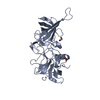 4rpbS S: Starting model for refinement |
|---|---|
| Similar structure data |


-Links
 PDBj
PDBj
- Assembly
| Deposited unit | 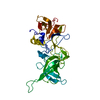
| ||||||||
|---|---|---|---|---|---|---|---|---|---|
| 1 | 
| ||||||||
| Unit cell |
| ||||||||
| Components on special symmetry positions |
|
-Components
| #1: Protein | Mass: 34149.219 Da / Num. of mol.: 1 Source method: isolated from a genetically manipulated source Source: (gene. exp.) Norovirus GIIProduction host: References: UniProt: A0A1X9W7M5, UniProt: A0A2R3BZ02*PLUS |
|---|---|
| #2: Water | ChemComp-HOH /  Mass: 18.015 Da / Num. of mol.: 421 / Source method: isolated from a natural source / Formula: H2O Mass: 18.015 Da / Num. of mol.: 421 / Source method: isolated from a natural source / Formula: H2O |
-Experimental details
-Experiment
| Experiment | Method: X-RAY DIFFRACTION / Number of used crystals: 1 |
|---|
- Sample preparation
| Crystal | Density Matthews: 2.53 Å3/Da / Density % sol: 51.45 % |
|---|---|
| Crystal grow | Temperature: 291 K / Method: evaporation / pH: 4.6 Details: 0.1 M Sodium Acetate trihydrate pH 4.6, 8% w/v Polyethylene Glycol 4000 |
-Data collection
| Diffraction | Mean temperature: 113 K |
|---|---|
| Diffraction source | Source: SYNCHROTRON / Site: SSRF / Beamline: BL17U1 / Wavelength: 0.97915 Å |
| Detector | Type: ADSC QUANTUM 315r / Detector: CCD / Date: Jul 3, 2017 |
| Radiation | Protocol: SINGLE WAVELENGTH / Monochromatic (M) / Laue (L): M / Scattering type: x-ray |
| Radiation wavelength | Wavelength: 0.97915 Å / Relative weight: 1 |
| Reflection | Resolution: 1.2→50 Å / Num. obs: 99924 / % possible obs: 99.7 % / Redundancy: 12.1 % / Net I/σ(I): 29.5 |
| Reflection shell | Resolution: 1.2→1.24 Å |
- Processing
| Software |
| ||||||||||||||||||||||||||||||||||||||||
|---|---|---|---|---|---|---|---|---|---|---|---|---|---|---|---|---|---|---|---|---|---|---|---|---|---|---|---|---|---|---|---|---|---|---|---|---|---|---|---|---|---|
| Refinement | Method to determine structure: MOLECULAR REPLACEMENT Starting model: 4RPB Resolution: 1.202→28.361 Å / SU ML: 0.1 / Cross valid method: FREE R-VALUE / σ(F): 1.35 / Phase error: 17.02
| ||||||||||||||||||||||||||||||||||||||||
| Solvent computation | Shrinkage radii: 0.9 Å / VDW probe radii: 1.11 Å | ||||||||||||||||||||||||||||||||||||||||
| Displacement parameters | Biso max: 115.06 Å2 / Biso mean: 24.0283 Å2 / Biso min: 8.63 Å2 | ||||||||||||||||||||||||||||||||||||||||
| Refinement step | Cycle: final / Resolution: 1.202→28.361 Å
| ||||||||||||||||||||||||||||||||||||||||
| Refine LS restraints |
| ||||||||||||||||||||||||||||||||||||||||
| LS refinement shell | Refine-ID: X-RAY DIFFRACTION / Rfactor Rfree error: 0 / % reflection obs: 100 %
| ||||||||||||||||||||||||||||||||||||||||
| Refinement TLS params. | Method: refined / Origin x: 25.8021 Å / Origin y: -38.8937 Å / Origin z: 10.3564 Å
| ||||||||||||||||||||||||||||||||||||||||
| Refinement TLS group | Selection details: all |
