 Movie
Movie Controller

[English] 日本語
 Yorodumi
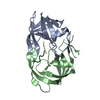
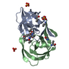
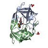
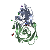
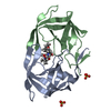
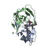
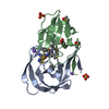
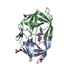
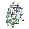
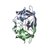
- PDB-5yrs: X-ray Snapshot of HIV-1 Protease in Action: Observation of Tetrah... -
+ Open data

- Basic information
| Entry | Database: PDB / ID: 5yrs | |||||||||
|---|---|---|---|---|---|---|---|---|---|---|
| Title | X-ray Snapshot of HIV-1 Protease in Action: Observation of Tetrahedral Intermediate and Its SIHB with Catalytic Aspartate | |||||||||
 Components Components |
| |||||||||
 Keywords Keywords | HYDROLASE / HIV-1 PROTEASE / TETRAHEDRAL INTERMEDIATE / SHORT IONIC HYDROGEN BOND / SUBSTRATE COMPLEX / RT-RH | |||||||||
| Function / homology |  Function and homology information Function and homology informationintegrase activity / Integration of viral DNA into host genomic DNA / Autointegration results in viral DNA circles / Minus-strand DNA synthesis / Plus-strand DNA synthesis / Uncoating of the HIV Virion / 2-LTR circle formation / Vpr-mediated nuclear import of PICs / Early Phase of HIV Life Cycle / Integration of provirus ...integrase activity / Integration of viral DNA into host genomic DNA / Autointegration results in viral DNA circles / Minus-strand DNA synthesis / Plus-strand DNA synthesis / Uncoating of the HIV Virion / 2-LTR circle formation / Vpr-mediated nuclear import of PICs / Early Phase of HIV Life Cycle / Integration of provirus / APOBEC3G mediated resistance to HIV-1 infection / Binding and entry of HIV virion / viral life cycle / HIV-1 retropepsin / symbiont-mediated activation of host apoptosis / retroviral ribonuclease H / exoribonuclease H / exoribonuclease H activity / protein processing / Assembly Of The HIV Virion / viral genome integration into host DNA / Budding and maturation of HIV virion / establishment of integrated proviral latency / RNA-directed DNA polymerase / RNA stem-loop binding / viral penetration into host nucleus / host multivesicular body / RNA-directed DNA polymerase activity / RNA-DNA hybrid ribonuclease activity / Transferases; Transferring phosphorus-containing groups; Nucleotidyltransferases / peptidase activity / host cell / viral nucleocapsid / DNA recombination / DNA-directed DNA polymerase / aspartic-type endopeptidase activity / Hydrolases; Acting on ester bonds / DNA-directed DNA polymerase activity / symbiont-mediated suppression of host gene expression / viral translational frameshifting / symbiont entry into host cell / lipid binding / host cell nucleus / host cell plasma membrane / virion membrane / structural molecule activity / DNA binding / zinc ion binding / identical protein binding Similarity search - Function | |||||||||
| Biological species |   Human immunodeficiency virus 1 Human immunodeficiency virus 1 | |||||||||
| Method |  X-RAY DIFFRACTION / SYNCHROTRON / MOLECULAR REPLACEMENT / Resolution: 1.76 Å X-RAY DIFFRACTION / SYNCHROTRON / MOLECULAR REPLACEMENT / Resolution: 1.76 Å | |||||||||
 Authors Authors | Das, A. / Mahale, S. / Prashar, V. / Bihani, S. / Ferrer, J.-L. / Hosur, M.V. | |||||||||
| Funding support |  India, 1items India, 1items
| |||||||||
 Citation Citation | Journal: J. Am. Chem. Soc. / Year: 2010 Title: X-ray snapshot of HIV-1 protease in action: observation of tetrahedral intermediate and short ionic hydrogen bond SIHB with catalytic aspartate. Authors: Das, A. / Mahale, S. / Prashar, V. / Bihani, S. / Ferrer, J.L. / Hosur, M.V. | |||||||||
| History |
|
- Structure visualization
| Structure viewer | Molecule: MolmilJmol/JSmol |
|---|
- Downloads & links
-Download
| PDBx/mmCIF format | 5yrs.cif.gz | 60 KB | Display | PDBx/mmCIF format |
|---|---|---|---|---|
| PDB format | pdb5yrs.ent.gz | 43.4 KB | Display | PDB format |
| PDBx/mmJSON format | 5yrs.json.gz | Tree view | PDBx/mmJSON format | |
| Others |  Other downloads Other downloads |
-Validation report
| Arichive directory | https://data.pdbj.org/pub/pdb/validation_reports/yr/5yrsftp://data.pdbj.org/pub/pdb/validation_reports/yr/5yrs | HTTPS FTP |
|---|
-Related structure data
| Related structure data |  1lv1S S: Starting model for refinement |
|---|---|
| Similar structure data |


-Links
 PDBj
PDBj


- Assembly
| Deposited unit | 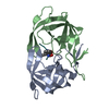
| ||||||||
|---|---|---|---|---|---|---|---|---|---|
| 1 |
| ||||||||
| Unit cell |
|
-Components
| #1: Protein | Mass: 11163.094 Da / Num. of mol.: 1 / Mutation: C95M Source method: isolated from a genetically manipulated source Source: (gene. exp.) Human immunodeficiency virus 1 / Strain: M:B_HXB2R / Gene: gag-pol, HIV-1 PR B / Plasmid: pET 11a / Production host:  |
|---|---|
| #2: Protein | Mass: 11102.976 Da / Num. of mol.: 1 / Mutation: C95A Source method: isolated from a genetically manipulated source Source: (gene. exp.) Human immunodeficiency virus 1 / Strain: M:B_HXB2R / Gene: gag-pol, HIV-1 PR B / Plasmid: pET 11a / Production host: |
| #3: Protein/peptide | Mass: 774.815 Da / Num. of mol.: 1 / Source method: obtained synthetically Details: THIS SEQUENCE CORRESPONDS TO RT-RH CLEAVAGE SITE, NATURALLY OCCURING IN HIV-1 GAG-POL POLYPROTEIN. Source: (synth.) Human immunodeficiency virus 1 / References: UniProt: P04585*PLUS |
| #4: Water | ChemComp-HOH /  Mass: 18.015 Da / Num. of mol.: 193 / Source method: isolated from a natural source / Formula: H2O Mass: 18.015 Da / Num. of mol.: 193 / Source method: isolated from a natural source / Formula: H2O |
| Has protein modification | Y |
| Sequence details | THE AUTHOR STATES THAT RESIDUE HPH X 4 IS L-PHENYLALANINE AND HYDRATED L-PHE RESIDUE HAVING ...THE AUTHOR STATES THAT RESIDUE HPH X 4 IS L-PHENYLALAN |
-Experimental details
-Experiment
| Experiment | Method: X-RAY DIFFRACTION / Number of used crystals: 1 |
|---|
- Sample preparation
| Crystal | Density Matthews: 1.99 Å3/Da / Density % sol: 38.16 % |
|---|---|
| Crystal grow | Temperature: 298 K / Method: vapor diffusion, hanging drop / pH: 6.2 Details: 0.1M PHOSPHATE-0.2M CITRATE BUFFER, AMM. SULPHATE, PH 6.2, VAPOR DIFFUSION, HANGING DROP, TEMPERATURE 298K |
-Data collection
| Diffraction | Mean temperature: 100 K |
|---|---|
| Diffraction source | Source: SYNCHROTRON / Site: ESRF  / Beamline: ID23-2 / Wavelength: 0.987 Å / Beamline: ID23-2 / Wavelength: 0.987 Å |
| Detector | Type: ADSC QUANTUM 315r / Detector: CCD / Date: Aug 5, 2008 |
| Radiation | Monochromator: A HORIZONTALLY DIFFRACTING SI (111) MONOCHROMATOR Protocol: SINGLE WAVELENGTH / Monochromatic (M) / Laue (L): M / Scattering type: x-ray |
| Radiation wavelength | Wavelength: 0.987 Å / Relative weight: 1 |
| Reflection | Resolution: 1.76→50 Å / Num. obs: 17226 / % possible obs: 95.3 % / Observed criterion σ(I): -3 / Redundancy: 7.5 % / Rmerge(I) obs: 0.072 / Net I/σ(I): 21.76 |
| Reflection shell | Resolution: 1.76→1.81 Å / Rmerge(I) obs: 0.465 / Mean I/σ(I) obs: 2.98 / % possible all: 88.5 |
- Processing
| Software |
| ||||||||||||||||||||||||||||||||||||||||||||||||||||||||||||
|---|---|---|---|---|---|---|---|---|---|---|---|---|---|---|---|---|---|---|---|---|---|---|---|---|---|---|---|---|---|---|---|---|---|---|---|---|---|---|---|---|---|---|---|---|---|---|---|---|---|---|---|---|---|---|---|---|---|---|---|---|---|
| Refinement | Method to determine structure: MOLECULAR REPLACEMENT Starting model: 1LV1 Resolution: 1.76→50 Å / Cross valid method: THROUGHOUT / σ(F): 2.5 / Stereochemistry target values: ENGH & HUBER
| ||||||||||||||||||||||||||||||||||||||||||||||||||||||||||||
| Refinement step | Cycle: 1 / Resolution: 1.76→50 Å
| ||||||||||||||||||||||||||||||||||||||||||||||||||||||||||||
| Refine LS restraints |
|
