 Movie
Movie Controller


+ Open data

- Basic information
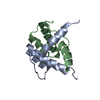
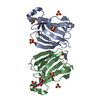
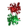
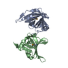
| Entry | Database: PDB / ID: 5yre | |||||||||||||||
|---|---|---|---|---|---|---|---|---|---|---|---|---|---|---|---|---|
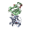
| Title | Crystal structure of PPL3A | |||||||||||||||
 Components Components | (PPL3-A) x 2 | |||||||||||||||
 Keywords Keywords | SUGAR BINDING PROTEIN / Lectin / Biomineralization / Post-translational modification / Calcite / Docking simulation | |||||||||||||||
| Function / homology | Jacalin-like lectin domain / Jacalin-type lectin domain profile. / Jacalin-like lectin domain / Jacalin-like lectin domain / Jacalin-like lectin domain superfamily / carbohydrate binding / Jacalin-related lectin Function and homology information Function and homology information | |||||||||||||||
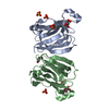
| Biological species |  Pteria penguin (invertebrata) Pteria penguin (invertebrata) | |||||||||||||||
| Method |  X-RAY DIFFRACTION / SYNCHROTRON / MOLECULAR REPLACEMENT / Resolution: 1.4 Å X-RAY DIFFRACTION / SYNCHROTRON / MOLECULAR REPLACEMENT / Resolution: 1.4 Å | |||||||||||||||
 Authors Authors | Nakae, S. / Shionyu, M. / Ogawa, T. / Shirai, T. | |||||||||||||||
| Funding support |  Japan, 4items Japan, 4items
| |||||||||||||||
 Citation Citation | Journal: Proteins / Year: 2018 Title: Structures of jacalin-related lectin PPL3 regulating pearl shell biomineralization Authors: Nakae, S. / Shionyu, M. / Ogawa, T. / Shirai, T. | |||||||||||||||
| History |
|
- Structure visualization
| Structure viewer | Molecule: MolmilJmol/JSmol |
|---|
- Downloads & links
-Download
| PDBx/mmCIF format | 5yre.cif.gz | 82.3 KB | Display | PDBx/mmCIF format |
|---|---|---|---|---|
| PDB format | pdb5yre.ent.gz | 59.3 KB | Display | PDB format |
| PDBx/mmJSON format | 5yre.json.gz | Tree view | PDBx/mmJSON format | |
| Others |  Other downloads Other downloads |
-Validation report
| Arichive directory | https://data.pdbj.org/pub/pdb/validation_reports/yr/5yreftp://data.pdbj.org/pub/pdb/validation_reports/yr/5yre | HTTPS FTP |
|---|
-Related structure data
| Related structure data |  5yrfC 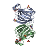 5yrgC  5yrhC  5yriC  5yrjC 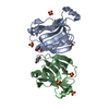 5yrkSC 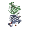 5yrlC 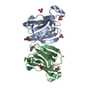 5yrmC S: Starting model for refinement C: citing same article ( |
|---|---|
| Similar structure data |


-Links
 PDBj
PDBj- Assembly
| Deposited unit | 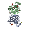
| ||||||||
|---|---|---|---|---|---|---|---|---|---|
| 1 |
| ||||||||
| Unit cell |
|
-Components
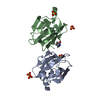
| #1: Protein | Mass: 15754.878 Da / Num. of mol.: 1 / Source method: isolated from a natural source / Source: (natural) Pteria penguin (invertebrata) / References: UniProt: B6F0T7 | ||||||||
|---|---|---|---|---|---|---|---|---|---|
| #2: Protein | Mass: 15722.835 Da / Num. of mol.: 1 / Source method: isolated from a natural source / Source: (natural) Pteria penguin (invertebrata) / References: UniProt: B6F0T7 | ||||||||
| #3: Chemical | ChemComp-SO4 /   Mass: 96.063 Da / Num. of mol.: 7 / Source method: obtained synthetically / Formula: SO4 Mass: 96.063 Da / Num. of mol.: 7 / Source method: obtained synthetically / Formula: SO4#4: Chemical |   Mass: 92.094 Da / Num. of mol.: 2 / Source method: obtained synthetically / Formula: C3H8O3 Mass: 92.094 Da / Num. of mol.: 2 / Source method: obtained synthetically / Formula: C3H8O3#5: Water | ChemComp-HOH / |  Mass: 18.015 Da / Num. of mol.: 418 / Source method: isolated from a natural source / Formula: H2O Mass: 18.015 Da / Num. of mol.: 418 / Source method: isolated from a natural source / Formula: H2OHas protein modification | Y | Sequence details | The N-terminus of subunit after signal peptide removal is Gln20-Val21 from mRNA, but is changed to ...The N-terminus of subunit after signal peptide removal is Gln20-Val21 from mRNA, but is changed to pGlu20-Val21 or Gln20-Ile21 because of post-translational modifications. The genebank accession for PPL3-a is AB425240.2. | |
-Experimental details
-Experiment
| Experiment | Method: X-RAY DIFFRACTION / Number of used crystals: 1 |
|---|
- Sample preparation
| Crystal | Density Matthews: 1.93 Å3/Da / Density % sol: 36.4 % |
|---|---|
| Crystal grow | Temperature: 293 K / Method: small tubes Details: 0.2M ammonium sulfate, 25%(w/v) PEG 3350, 0.1M Tris/bis-Tris buffer pH 5.5 microgravity environments on ISS |
-Data collection
| Diffraction | Mean temperature: 100 K |
|---|---|
| Diffraction source | Source: SYNCHROTRON / Site: SPring-8 / Beamline: BL26B1 / Wavelength: 1 Å |
| Detector | Type: RAYONIX MX225HE / Detector: CCD / Date: Nov 19, 2016 |
| Radiation | Protocol: SINGLE WAVELENGTH / Monochromatic (M) / Laue (L): M / Scattering type: x-ray |
| Radiation wavelength | Wavelength: 1 Å / Relative weight: 1 |
| Reflection | Resolution: 1.4→20 Å / Num. obs: 44177 / % possible obs: 90.3 % / Redundancy: 2.7 % / Rmerge(I) obs: 0.016 / Net I/σ(I): 27.1 |
| Reflection shell | Resolution: 1.4→1.48 Å / Redundancy: 2.8 % / Rmerge(I) obs: 0.148 / Mean I/σ(I) obs: 5.2 / Num. unique obs: 5714 / % possible all: 81.4 |
- Processing
| Software |
| |||||||||||||||||||||||||||||||||||||||||||||||||||||||||||||||||||||||||||||||||||||||||||||||||||||||||||||||||||||||
|---|---|---|---|---|---|---|---|---|---|---|---|---|---|---|---|---|---|---|---|---|---|---|---|---|---|---|---|---|---|---|---|---|---|---|---|---|---|---|---|---|---|---|---|---|---|---|---|---|---|---|---|---|---|---|---|---|---|---|---|---|---|---|---|---|---|---|---|---|---|---|---|---|---|---|---|---|---|---|---|---|---|---|---|---|---|---|---|---|---|---|---|---|---|---|---|---|---|---|---|---|---|---|---|---|---|---|---|---|---|---|---|---|---|---|---|---|---|---|---|---|
| Refinement | Method to determine structure: MOLECULAR REPLACEMENT Starting model: 5YRK Resolution: 1.4→19.739 Å / SU ML: 0.17 / Cross valid method: FREE R-VALUE / σ(F): 1.33 / Phase error: 21.72
| |||||||||||||||||||||||||||||||||||||||||||||||||||||||||||||||||||||||||||||||||||||||||||||||||||||||||||||||||||||||
| Solvent computation | Shrinkage radii: 0.9 Å / VDW probe radii: 1.11 Å | |||||||||||||||||||||||||||||||||||||||||||||||||||||||||||||||||||||||||||||||||||||||||||||||||||||||||||||||||||||||
| Refinement step | Cycle: LAST / Resolution: 1.4→19.739 Å
| |||||||||||||||||||||||||||||||||||||||||||||||||||||||||||||||||||||||||||||||||||||||||||||||||||||||||||||||||||||||
| Refine LS restraints |
| |||||||||||||||||||||||||||||||||||||||||||||||||||||||||||||||||||||||||||||||||||||||||||||||||||||||||||||||||||||||
| LS refinement shell |
|
