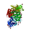
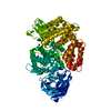
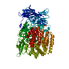
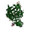
[English] 日本語
Yorodumi
- PDB-5yo1: Structure of ePepN E298A mutant in complex with Puromycin -

+
Open data


ID or keywords:

Loading...

-
Basic information

Entry
Database: PDB / ID: 5yo1
TitleStructure of ePepN E298A mutant in complex with Puromycin
ComponentsAminopeptidase N
KeywordsHYDROLASE / M1 class aminopeptidase
Function / homology
Function and homology information


membrane alanyl aminopeptidase / aminopeptidase activity / metallopeptidase activity / proteolysis / zinc ion binding / identical protein binding / plasma membrane
Similarity search - Function
Peptidase M1, alanyl aminopeptidase, C-terminal domain / Aminopeptidase N, middle-beta domain / Peptidase M1, alanyl aminopeptidase / Peptidase M1, alanyl aminopeptidase, C-terminal / Peptidase M1, alanyl aminopeptidase, Ig-like fold / Peptidase M1, alanyl aminopeptidase, C-terminal domain superfamily / Alanyl aminopeptidase, Ig-like domain superfamily / Domain of unknown function (DUF3458) Ig-like fold / Domain of unknown function (DUF3458_C) ARM repeats / Zincin-like fold ...Peptidase M1, alanyl aminopeptidase, C-terminal domain / Aminopeptidase N, middle-beta domain / Peptidase M1, alanyl aminopeptidase / Peptidase M1, alanyl aminopeptidase, C-terminal / Peptidase M1, alanyl aminopeptidase, Ig-like fold / Peptidase M1, alanyl aminopeptidase, C-terminal domain superfamily / Alanyl aminopeptidase, Ig-like domain superfamily / Domain of unknown function (DUF3458) Ig-like fold / Domain of unknown function (DUF3458_C) ARM repeats / Zincin-like fold / Zincin-like - #30 / Zincin-like / tricorn interacting facor f3 domain / Aminopeptidase N-like , N-terminal domain / Peptidase M1, alanine aminopeptidase/leukotriene A4 hydrolase / Peptidase M1, membrane alanine aminopeptidase / Peptidase family M1 domain / Peptidase M1 N-terminal domain / Aminopeptidase N-like , N-terminal domain superfamliy / Neutral Protease Domain 2 / Neutral Protease; domain 2 / Peptidase M4/M1, CTD superfamily / Neutral zinc metallopeptidases, zinc-binding region signature. / Alpha Horseshoe / Immunoglobulin-like / Sandwich / 2-Layer Sandwich / Orthogonal Bundle / Mainly Beta / Mainly Alpha / Alpha Beta
Similarity search - Domain/homology
PUROMYCIN / Aminopeptidase N
Similarity search - Component
Biological speciesEscherichia coli K12 (bacteria)
MethodX-RAY DIFFRACTION / MOLECULAR REPLACEMENT / Resolution: 2.5 Å
AuthorsGanji, R.J. / Reddi, R. / Marapaka, A.K. / Addlagatta, A.
Funding support India, 1items
OrganizationGrant numberCountry
BT-BRB-TF-2-2011 India
CitationJournal: Int.J.Biol.Macromol. / Year: 2020
Title: Puromycin, a selective inhibitor of PSA acts as a substrate for other M1 family aminopeptidases: Biochemical and structural basis
Authors: Reddi, R. / Ganji, R.J. / Marapaka, A.K. / Bala, S.C. / Yerra, N.V. / Haque, N. / Addlagatta, A.
History
DepositionOct 26, 2017Deposition site: PDBJ / Processing site: PDBJ
Revision 1.0Nov 7, 2018Provider: repository / Type: Initial release
Revision 1.1Jul 27, 2022Group: Database references / Category: citation / citation_author / database_2
Item: _citation.country / _citation.journal_abbrev ..._citation.country / _citation.journal_abbrev / _citation.journal_id_ASTM / _citation.journal_id_CSD / _citation.journal_id_ISSN / _citation.journal_volume / _citation.page_first / _citation.page_last / _citation.pdbx_database_id_DOI / _citation.title / _citation.year / _database_2.pdbx_DOI / _database_2.pdbx_database_accession
Revision 1.2Nov 22, 2023Group: Data collection / Refinement description
Category: chem_comp_atom / chem_comp_bond / pdbx_initial_refinement_model

-
Structure visualization

Structure viewerMolecule:
MolmilJmol/JSmol

Downloads & links

-
Assembly

Deposited unit
A: Aminopeptidase N
hetero molecules


Theoretical massNumber of molelcules
Total (without water)103,52017
Polymers101,3831
Non-polymers2,13716
Water17,781987
1


  • Idetical with deposited unit
  • defined by author&software
  • Evidence: gel filtration
TypeNameSymmetry operationNumber
identity operation1_555x,y,z1
Buried area100 Å2
ΔGint-37 kcal/mol
Surface area32670 Å2
MethodPISA
Unit cell
Length a, b, c (Å)120.325, 120.325, 170.012
Angle α, β, γ (deg.)90.00, 90.00, 120.00
Int Tables number152
Space group name H-MP3121

-
Components

-
Protein , 1 types, 1 molecules A

#1: Protein Aminopeptidase N / Alpha-aminoacylpeptide hydrolase / E. coli Amino peptidase N


Mass: 101383.156 Da / Num. of mol.: 1 / Mutation: E298A
Source method: isolated from a genetically manipulated source
Source: (gene. exp.) Escherichia coli K12 (bacteria) / Strain: K12 / Gene: pepN, b0932, JW0915 / Production host: Escherichia coli (E. coli) / References: UniProt: P04825, membrane alanyl aminopeptidase

-
Non-polymers , 5 types, 1003 molecules

#2: Chemical ChemComp-ZN / ZINC ION


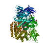
Mass: 65.409 Da / Num. of mol.: 1 / Source method: obtained synthetically / Formula: Zn
#3: Chemical ChemComp-PUY / PUROMYCIN


Type: RNA linking / Mass: 471.510 Da / Num. of mol.: 2 / Source method: obtained synthetically / Formula: C22H29N7O5 / Feature type: SUBJECT OF INVESTIGATION / Comment: antibiotic, inhibitor*YM
#4: Chemical ChemComp-NA / SODIUM ION


Mass: 22.990 Da / Num. of mol.: 1 / Source method: obtained synthetically / Formula: Na
#5: Chemical
ChemComp-GOL / GLYCEROL / GLYCERIN / PROPANE-1,2,3-TRIOL


Mass: 92.094 Da / Num. of mol.: 12 / Source method: obtained synthetically / Formula: C3H8O3
#6: Water ChemComp-HOH / water


Mass: 18.015 Da / Num. of mol.: 987 / Source method: isolated from a natural source / Formula: H2O

-
Experimental details

-
Experiment

ExperimentMethod: X-RAY DIFFRACTION / Number of used crystals: 1

-
Sample preparation

CrystalDensity Matthews: 3.5 Å3/Da / Density % sol: 64.9 %
Crystal growTemperature: 298 K / Method: vapor diffusion, hanging drop / pH: 7.5 / Details: 2.0M Sodium Malonate

-
Data collection

DiffractionMean temperature: 100 K
Diffraction sourceSource: ROTATING ANODE / Type: RIGAKU MICROMAX-007 HF / Wavelength: 1.54 Å
DetectorType: RIGAKU RAXIS IV++ / Detector: IMAGE PLATE / Date: Apr 20, 2015
RadiationProtocol: SINGLE WAVELENGTH / Monochromatic (M) / Laue (L): M / Scattering type: x-ray
Radiation wavelengthWavelength: 1.54 Å / Relative weight: 1
ReflectionResolution: 2.5→19.9 Å / Num. obs: 49464 / % possible obs: 99 % / Redundancy: 4.3 % / Net I/σ(I): 2.13

-
Processing

Software
NameVersionClassification
REFMAC5.8.0155refinement
HKL-3000data reduction
HKL-3000data scaling
MOLREPphasing
RefinementMethod to determine structure: MOLECULAR REPLACEMENT
Starting model: 2HPO
Resolution: 2.5→19.9 Å / Cor.coef. Fo:Fc: 0.974 / Cor.coef. Fo:Fc free: 0.947 / SU B: 6.187 / SU ML: 0.133 / Cross valid method: THROUGHOUT / ESU R: 0.238 / ESU R Free: 0.195 / Details: HYDROGENS HAVE BEEN ADDED IN THE RIDING POSITIONS
RfactorNum. reflection% reflectionSelection details
Rfree0.18427 2503 5.1 %RANDOM
Rwork0.1258 ---
obs0.12876 46961 99.41 %-
Solvent computationIon probe radii: 0.8 Å / Shrinkage radii: 0.8 Å / VDW probe radii: 1.2 Å
Displacement parametersBiso mean: 33.438 Å2
Baniso -1Baniso -2Baniso -3
1--0 Å2-0 Å20 Å2
2---0 Å2-0 Å2
3---0.01 Å2
Refinement stepCycle: 1 / Resolution: 2.5→19.9 Å
ProteinNucleic acidLigandSolventTotal
Num. atoms6936 0 142 987 8065
Refine LS restraints
Refine-IDTypeDev idealDev ideal targetNumber
X-RAY DIFFRACTIONr_bond_refined_d0.0160.0197225
X-RAY DIFFRACTIONr_bond_other_d0.0020.026797
X-RAY DIFFRACTIONr_angle_refined_deg1.8541.9679798
X-RAY DIFFRACTIONr_angle_other_deg1.052315583
X-RAY DIFFRACTIONr_dihedral_angle_1_deg6.4555865
X-RAY DIFFRACTIONr_dihedral_angle_2_deg38.05124.176364
X-RAY DIFFRACTIONr_dihedral_angle_3_deg15.064151187
X-RAY DIFFRACTIONr_dihedral_angle_4_deg19.5541553
X-RAY DIFFRACTIONr_chiral_restr0.1060.21087
X-RAY DIFFRACTIONr_gen_planes_refined0.0080.0218194
X-RAY DIFFRACTIONr_gen_planes_other0.0020.021719
X-RAY DIFFRACTIONr_nbd_refined
X-RAY DIFFRACTIONr_nbd_other
X-RAY DIFFRACTIONr_nbtor_refined
X-RAY DIFFRACTIONr_nbtor_other
X-RAY DIFFRACTIONr_xyhbond_nbd_refined
X-RAY DIFFRACTIONr_xyhbond_nbd_other
X-RAY DIFFRACTIONr_metal_ion_refined
X-RAY DIFFRACTIONr_metal_ion_other
X-RAY DIFFRACTIONr_symmetry_vdw_refined
X-RAY DIFFRACTIONr_symmetry_vdw_other
X-RAY DIFFRACTIONr_symmetry_hbond_refined
X-RAY DIFFRACTIONr_symmetry_hbond_other
X-RAY DIFFRACTIONr_symmetry_metal_ion_refined
X-RAY DIFFRACTIONr_symmetry_metal_ion_other
X-RAY DIFFRACTIONr_mcbond_it2.3162.9273469
X-RAY DIFFRACTIONr_mcbond_other2.3162.9273469
X-RAY DIFFRACTIONr_mcangle_it3.5244.384331
X-RAY DIFFRACTIONr_mcangle_other3.5234.3824332
X-RAY DIFFRACTIONr_scbond_it3.7453.4093756
X-RAY DIFFRACTIONr_scbond_other3.7423.4093756
X-RAY DIFFRACTIONr_scangle_it
X-RAY DIFFRACTIONr_scangle_other5.7794.9315468
X-RAY DIFFRACTIONr_long_range_B_refined34.86940.6849074
X-RAY DIFFRACTIONr_long_range_B_other34.86740.6919075
X-RAY DIFFRACTIONr_rigid_bond_restr
X-RAY DIFFRACTIONr_sphericity_free
X-RAY DIFFRACTIONr_sphericity_bonded
LS refinement shellResolution: 2.502→2.566 Å / Total num. of bins used: 20
RfactorNum. reflection% reflection
Rfree0.276 199 -
Rwork0.208 3383 -
obs--99.42 %

+
About Yorodumi

-
News

-
Feb 9, 2022. New format data for meta-information of EMDB entries

New format data for meta-information of EMDB entries

  • Version 3 of the EMDB header file is now the official format.
  • The previous official version 1.9 will be removed from the archive.

Related info.:EMDB header

External links:wwPDB to switch to version 3 of the EMDB data model

-
Aug 12, 2020. Covid-19 info

Covid-19 info

URL: https://pdbjlc1.pdbj.org/emnavi/covid19.php

New page: Covid-19 featured information page in EM Navigator.

Related info.:Covid-19 info / Mar 5, 2020. Novel coronavirus structure data

+
Mar 5, 2020. Novel coronavirus structure data

Novel coronavirus structure data

Related info.:Yorodumi Speices / Aug 12, 2020. Covid-19 info

External links:COVID-19 featured content - PDBj / Molecule of the Month (242):Coronavirus Proteases

+
Jan 31, 2019. EMDB accession codes are about to change! (news from PDBe EMDB page)

EMDB accession codes are about to change! (news from PDBe EMDB page)

  • The allocation of 4 digits for EMDB accession codes will soon come to an end. Whilst these codes will remain in use, new EMDB accession codes will include an additional digit and will expand incrementally as the available range of codes is exhausted. The current 4-digit format prefixed with “EMD-” (i.e. EMD-XXXX) will advance to a 5-digit format (i.e. EMD-XXXXX), and so on. It is currently estimated that the 4-digit codes will be depleted around Spring 2019, at which point the 5-digit format will come into force.
  • The EM Navigator/Yorodumi systems omit the EMD- prefix.

Related info.:Q: What is EMD? / ID/Accession-code notation in Yorodumi/EM Navigator

External links:EMDB Accession Codes are Changing Soon! / Contact to PDBj

+
Jul 12, 2017. Major update of PDB

Major update of PDB

  • wwPDB released updated PDB data conforming to the new PDBx/mmCIF dictionary.
  • This is a major update changing the version number from 4 to 5, and with Remediation, in which all the entries are updated.
  • In this update, many items about electron microscopy experimental information are reorganized (e.g. em_software).
  • Now, EM Navigator and Yorodumi are based on the updated data.

External links:wwPDB Remediation / Enriched Model Files Conforming to OneDep Data Standards Now Available in the PDB FTP Archive

-
Yorodumi

Thousand views of thousand structures

  • Yorodumi is a browser for structure data from EMDB, PDB, SASBDB, etc.
  • This page is also the successor to EM Navigator detail page, and also detail information page/front-end page for Omokage search.
  • The word "yorodu" (or yorozu) is an old Japanese word meaning "ten thousand". "mi" (miru) is to see.

Related info.:EMDB / PDB / SASBDB / Comparison of 3 databanks / Yorodumi Search / Aug 31, 2016. New EM Navigator & Yorodumi / Yorodumi Papers / Jmol/JSmol / Function and homology information / Changes in new EM Navigator and Yorodumi

Read more