| Entry | Database: PDB / ID: 4q4e
|
|---|
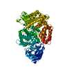
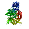
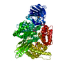
| Title | Crystal structure of E.coli aminopeptidase N in complex with actinonin |
|---|
 Components Components | Aminopeptidase N |
|---|
 Keywords Keywords | HYDROLASE / aminopeptidase |
|---|
| Function / homology |  Function and homology information Function and homology information
membrane alanyl aminopeptidase / alanyl aminopeptidase activity / aminopeptidase activity / metallopeptidase activity / proteolysis / zinc ion binding / identical protein binding / plasma membraneSimilarity search - Function Aminopeptidase N, middle-beta domain / Peptidase M1, alanyl aminopeptidase, C-terminal domain / Peptidase M1, alanyl aminopeptidase / Peptidase M1, alanyl aminopeptidase, C-terminal / Peptidase M1, alanyl aminopeptidase, Ig-like fold / Peptidase M1, alanyl aminopeptidase, C-terminal domain superfamily / Alanyl aminopeptidase, Ig-like domain superfamily / Domain of unknown function (DUF3458) Ig-like fold / Domain of unknown function (DUF3458_C) ARM repeats / Zincin-like fold ...Aminopeptidase N, middle-beta domain / Peptidase M1, alanyl aminopeptidase, C-terminal domain / Peptidase M1, alanyl aminopeptidase / Peptidase M1, alanyl aminopeptidase, C-terminal / Peptidase M1, alanyl aminopeptidase, Ig-like fold / Peptidase M1, alanyl aminopeptidase, C-terminal domain superfamily / Alanyl aminopeptidase, Ig-like domain superfamily / Domain of unknown function (DUF3458) Ig-like fold / Domain of unknown function (DUF3458_C) ARM repeats / Zincin-like fold / Zincin-like - #30 / Zincin-like / tricorn interacting facor f3 domain / Peptidase M1, alanine aminopeptidase/leukotriene A4 hydrolase / Peptidase M1, membrane alanine aminopeptidase / Aminopeptidase N-like , N-terminal domain / Peptidase family M1 domain / Peptidase M1 N-terminal domain / Aminopeptidase N-like , N-terminal domain superfamliy / Neutral Protease Domain 2 / Neutral Protease; domain 2 / Peptidase M4/M1, CTD superfamily / Neutral zinc metallopeptidases, zinc-binding region signature. / Alpha Horseshoe / Immunoglobulin-like / Sandwich / 2-Layer Sandwich / Orthogonal Bundle / Mainly Beta / Mainly Alpha / Alpha BetaSimilarity search - Domain/homology |
|---|
| Biological species |   Escherichia coli (E. coli) Escherichia coli (E. coli) |
|---|
| Method |  X-RAY DIFFRACTION / MOLECULAR REPLACEMENT / Resolution: 1.9 Å X-RAY DIFFRACTION / MOLECULAR REPLACEMENT / Resolution: 1.9 Å |
|---|
 Authors Authors | Reddi, R. / Ganji, R.J. / Addlagatta, A. |
|---|
 Citation Citation | Journal: Protein Sci. / Year: 2015
Title: Structural basis for the inhibition of M1 family aminopeptidases by the natural product actinonin: Crystal structure in complex with E. coli aminopeptidase N.
Authors: Ganji, R.J. / Reddi, R. / Gumpena, R. / Marapaka, A.K. / Arya, T. / Sankoju, P. / Bhukya, S. / Addlagatta, A. |
|---|
| History | | Deposition | Apr 14, 2014 | Deposition site: RCSB / Processing site: PDBJ |
|---|
| Revision 1.0 | Apr 15, 2015 | Provider: repository / Type: Initial release |
|---|
| Revision 1.1 | Dec 4, 2019 | Group: Data collection / Database references / Category: reflns_shell / struct_ref_seq_dif / Item: _struct_ref_seq_dif.details |
|---|
| Revision 1.2 | Aug 24, 2022 | Group: Database references / Derived calculations
Category: citation / database_2 ...citation / database_2 / pdbx_struct_conn_angle / struct_conn / struct_site
Item: _citation.journal_volume / _citation.page_first ..._citation.journal_volume / _citation.page_first / _citation.page_last / _citation.title / _database_2.pdbx_DOI / _database_2.pdbx_database_accession / _pdbx_struct_conn_angle.ptnr1_auth_comp_id / _pdbx_struct_conn_angle.ptnr1_auth_seq_id / _pdbx_struct_conn_angle.ptnr1_label_asym_id / _pdbx_struct_conn_angle.ptnr1_label_atom_id / _pdbx_struct_conn_angle.ptnr1_label_comp_id / _pdbx_struct_conn_angle.ptnr1_label_seq_id / _pdbx_struct_conn_angle.ptnr2_auth_comp_id / _pdbx_struct_conn_angle.ptnr2_auth_seq_id / _pdbx_struct_conn_angle.ptnr2_label_asym_id / _pdbx_struct_conn_angle.ptnr2_label_atom_id / _pdbx_struct_conn_angle.ptnr2_label_comp_id / _pdbx_struct_conn_angle.ptnr3_auth_comp_id / _pdbx_struct_conn_angle.ptnr3_auth_seq_id / _pdbx_struct_conn_angle.ptnr3_label_asym_id / _pdbx_struct_conn_angle.ptnr3_label_atom_id / _pdbx_struct_conn_angle.ptnr3_label_comp_id / _pdbx_struct_conn_angle.ptnr3_label_seq_id / _pdbx_struct_conn_angle.value / _struct_conn.pdbx_dist_value / _struct_conn.ptnr1_auth_comp_id / _struct_conn.ptnr1_auth_seq_id / _struct_conn.ptnr1_label_asym_id / _struct_conn.ptnr1_label_atom_id / _struct_conn.ptnr1_label_comp_id / _struct_conn.ptnr1_label_seq_id / _struct_conn.ptnr2_auth_comp_id / _struct_conn.ptnr2_auth_seq_id / _struct_conn.ptnr2_label_asym_id / _struct_conn.ptnr2_label_atom_id / _struct_conn.ptnr2_label_comp_id / _struct_site.pdbx_auth_asym_id / _struct_site.pdbx_auth_comp_id / _struct_site.pdbx_auth_seq_id |
|---|
| Revision 1.3 | May 29, 2024 | Group: Data collection / Category: chem_comp_atom / chem_comp_bond |
|---|
|
|---|
 Movie
Movie Controller

 Yorodumi
 Open data
 Basic information
 Structure visualization
 Downloads & links
 Other downloads


 PDBj
PDBj Assembly
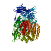


 Mass: 65.409 Da / Num. of mol.: 1 / Source method: obtained synthetically / Formula: Zn
 Mass: 385.498 Da / Num. of mol.: 1 / Source method: obtained synthetically / Formula: C19H35N3O5 / Comment: antitumor, antibiotic*YM
 Mass: 92.094 Da / Num. of mol.: 8 / Source method: obtained synthetically / Formula: C3H8O3
 Mass: 22.990 Da / Num. of mol.: 1 / Source method: obtained synthetically / Formula: Na
 Sample preparation
 Processing