 Movie
Movie Controller

[English] 日本語
 Yorodumi
- PDB-5ykd: Crystal structure of dihydropyrimidinase from Pseudomonas aerugin... -
+ Open data

- Basic information
| Entry | Database: PDB / ID: 5ykd | ||||||
|---|---|---|---|---|---|---|---|
| Title | Crystal structure of dihydropyrimidinase from Pseudomonas aeruginosa PAO1 at 2.17 angstrom resolution | ||||||
 Components Components | D-hydantoinase/dihydropyrimidinase | ||||||
 Keywords Keywords | HYDROLASE / dihydropyrimidinase | ||||||
| Function / homology |  Function and homology information Function and homology informationdihydropyrimidinase / pyrimidine-containing compound metabolic process / dihydropyrimidinase activity / hydrolase activity, acting on carbon-nitrogen (but not peptide) bonds, in cyclic amides / nucleobase-containing small molecule metabolic process / metal ion binding / cytosol Similarity search - Function | ||||||
| Biological species |   Pseudomonas aeruginosa (bacteria) Pseudomonas aeruginosa (bacteria) | ||||||
| Method |  X-RAY DIFFRACTION / SYNCHROTRON / MOLECULAR REPLACEMENT / Resolution: 2.17 Å X-RAY DIFFRACTION / SYNCHROTRON / MOLECULAR REPLACEMENT / Resolution: 2.17 Å | ||||||
 Authors Authors | Huang, Y.H. / Huang, C.Y. | ||||||
 Citation Citation | Journal: Bioinorg Chem Appl / Year: 2018 Title: Structural Basis for pH-Dependent Oligomerization of Dihydropyrimidinase fromPseudomonas aeruginosaPAO1. Authors: Cheng, J.H. / Huang, C.C. / Huang, Y.H. / Huang, C.Y. | ||||||
| History |
|
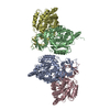
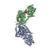
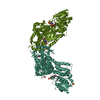
- Structure visualization
| Structure viewer | Molecule: MolmilJmol/JSmol |
|---|
- Downloads & links
-Download
| PDBx/mmCIF format | 5ykd.cif.gz | 370.5 KB | Display | PDBx/mmCIF format |
|---|---|---|---|---|
| PDB format | pdb5ykd.ent.gz | 301.8 KB | Display | PDB format |
| PDBx/mmJSON format | 5ykd.json.gz | Tree view | PDBx/mmJSON format | |
| Others |  Other downloads Other downloads |
-Validation report
| Arichive directory | https://data.pdbj.org/pub/pdb/validation_reports/yk/5ykdftp://data.pdbj.org/pub/pdb/validation_reports/yk/5ykd | HTTPS FTP |
|---|
-Related structure data
| Related structure data |  5e5cS S: Starting model for refinement |
|---|---|
| Similar structure data |


-Links
 PDBj
PDBj

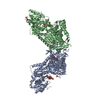
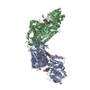
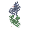
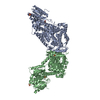
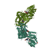
- Assembly
| Deposited unit | 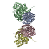
| ||||||||
|---|---|---|---|---|---|---|---|---|---|
| 1 |
| ||||||||
| Unit cell |
|
-Components
| #1: Protein | Mass: 52315.844 Da / Num. of mol.: 4 Source method: isolated from a genetically manipulated source Source: (gene. exp.) Pseudomonas aeruginosa (strain ATCC 15692 / DSM 22644 / CIP 104116 / JCM 14847 / LMG 12228 / 1C / PRS 101 / PAO1) (bacteria)Strain: ATCC 15692 / DSM 22644 / CIP 104116 / JCM 14847 / LMG 12228 / 1C / PRS 101 / PAO1 Gene: dht, PA0441 / Production host: #2: Chemical | ChemComp-ZN /   Mass: 65.409 Da / Num. of mol.: 8 / Source method: obtained synthetically / Formula: Zn Mass: 65.409 Da / Num. of mol.: 8 / Source method: obtained synthetically / Formula: Zn#3: Water | ChemComp-HOH / |  Mass: 18.015 Da / Num. of mol.: 313 / Source method: isolated from a natural source / Formula: H2O Mass: 18.015 Da / Num. of mol.: 313 / Source method: isolated from a natural source / Formula: H2O |
|---|
-Experimental details
-Experiment
| Experiment | Method: X-RAY DIFFRACTION / Number of used crystals: 1 |
|---|
- Sample preparation
| Crystal | Density Matthews: 2.32 Å3/Da / Density % sol: 46.94 % |
|---|---|
| Crystal grow | Temperature: 298 K / Method: vapor diffusion, hanging drop / pH: 5.9 Details: 10% PEG 8000, 100 mM HEPES, 200 mM calcium acetate pH 5.9 |
-Data collection
| Diffraction | Mean temperature: 298 K |
|---|---|
| Diffraction source | Source: SYNCHROTRON / Site: NSRRC  / Beamline: BL13C1 / Wavelength: 0.975 Å / Beamline: BL13C1 / Wavelength: 0.975 Å |
| Detector | Type: RAYONIX MX300HE / Detector: CCD / Date: Dec 17, 2015 |
| Radiation | Protocol: SINGLE WAVELENGTH / Monochromatic (M) / Laue (L): M / Scattering type: x-ray |
| Radiation wavelength | Wavelength: 0.975 Å / Relative weight: 1 |
| Reflection | Resolution: 2.17→117.79 Å / Num. obs: 100197 / % possible obs: 99.82 % / Redundancy: 7.1 % / Rmerge(I) obs: 0.122 / Net I/σ(I): 15.13 |
| Reflection shell | Resolution: 2.17→2.26 Å / Redundancy: 7.3 % / Rmerge(I) obs: 0.599 / Mean I/σ(I) obs: 3.7 / % possible all: 100 |
- Processing
| Software |
| ||||||||||||||||||||||||||||||||||||||||||||||||||||||||||||||||||||||||||||||||||||||||||||||||||||||||||||||||||||||||||||||||||||||||||||||||||||||||||||||||||||||||||||||||||||||
|---|---|---|---|---|---|---|---|---|---|---|---|---|---|---|---|---|---|---|---|---|---|---|---|---|---|---|---|---|---|---|---|---|---|---|---|---|---|---|---|---|---|---|---|---|---|---|---|---|---|---|---|---|---|---|---|---|---|---|---|---|---|---|---|---|---|---|---|---|---|---|---|---|---|---|---|---|---|---|---|---|---|---|---|---|---|---|---|---|---|---|---|---|---|---|---|---|---|---|---|---|---|---|---|---|---|---|---|---|---|---|---|---|---|---|---|---|---|---|---|---|---|---|---|---|---|---|---|---|---|---|---|---|---|---|---|---|---|---|---|---|---|---|---|---|---|---|---|---|---|---|---|---|---|---|---|---|---|---|---|---|---|---|---|---|---|---|---|---|---|---|---|---|---|---|---|---|---|---|---|---|---|---|---|
| Refinement | Method to determine structure: MOLECULAR REPLACEMENT Starting model: 5E5C Resolution: 2.17→30 Å / Cor.coef. Fo:Fc: 0.965 / Cor.coef. Fo:Fc free: 0.939 / SU B: 6.011 / SU ML: 0.152 / Cross valid method: THROUGHOUT / ESU R: 0.238 / ESU R Free: 0.198 / Details: HYDROGENS HAVE BEEN ADDED IN THE RIDING POSITIONS
| ||||||||||||||||||||||||||||||||||||||||||||||||||||||||||||||||||||||||||||||||||||||||||||||||||||||||||||||||||||||||||||||||||||||||||||||||||||||||||||||||||||||||||||||||||||||
| Solvent computation | Ion probe radii: 0.8 Å / Shrinkage radii: 0.8 Å / VDW probe radii: 1.2 Å | ||||||||||||||||||||||||||||||||||||||||||||||||||||||||||||||||||||||||||||||||||||||||||||||||||||||||||||||||||||||||||||||||||||||||||||||||||||||||||||||||||||||||||||||||||||||
| Displacement parameters | Biso mean: 39.431 Å2
| ||||||||||||||||||||||||||||||||||||||||||||||||||||||||||||||||||||||||||||||||||||||||||||||||||||||||||||||||||||||||||||||||||||||||||||||||||||||||||||||||||||||||||||||||||||||
| Refinement step | Cycle: 1 / Resolution: 2.17→30 Å
| ||||||||||||||||||||||||||||||||||||||||||||||||||||||||||||||||||||||||||||||||||||||||||||||||||||||||||||||||||||||||||||||||||||||||||||||||||||||||||||||||||||||||||||||||||||||
| Refine LS restraints |
|