 Movie
Movie Controller


+ Open data

- Basic information
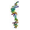
| Entry | Database: PDB / ID: 5yk7 | ||||||
|---|---|---|---|---|---|---|---|
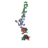
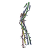
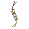
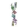
| Title | Crystal Structure of Mdm12-Mmm1 complex | ||||||
 Components Components |
| ||||||
 Keywords Keywords | LIPID TRANSPORT / Mmm1 / Mdm12 / ERMES / Phospholipid / Membrane contact site | ||||||
| Function / homology |  Function and homology information Function and homology informationERMES complex / mitochondrion inheritance / outer mitochondrial membrane protein complex / mitochondrial outer membrane translocase complex assembly / lipid transfer activity / aminophospholipid transport / peroxisome organization / protein insertion into mitochondrial outer membrane / mitochondria-associated endoplasmic reticulum membrane contact site / phospholipid transport ...ERMES complex / mitochondrion inheritance / outer mitochondrial membrane protein complex / mitochondrial outer membrane translocase complex assembly / lipid transfer activity / aminophospholipid transport / peroxisome organization / protein insertion into mitochondrial outer membrane / mitochondria-associated endoplasmic reticulum membrane contact site / phospholipid transport / phospholipid homeostasis / lipid transport / : / mitochondrion organization / mitochondrial outer membrane / lipid binding / endoplasmic reticulum membrane / mitochondrion / nucleus / plasma membrane / cytoplasm Similarity search - Function | ||||||
| Biological species |  Zygosaccharomyces rouxii (yeast) Zygosaccharomyces rouxii (yeast) | ||||||
| Method |  X-RAY DIFFRACTION / SYNCHROTRON / MOLECULAR REPLACEMENT / Resolution: 3.799 Å X-RAY DIFFRACTION / SYNCHROTRON / MOLECULAR REPLACEMENT / Resolution: 3.799 Å | ||||||
 Authors Authors | Jeong, H. / Park, J. / Lee, C. | ||||||
 Citation Citation | Journal: Proc. Natl. Acad. Sci. U.S.A. / Year: 2017 Title: Crystal structures of Mmm1 and Mdm12-Mmm1 reveal mechanistic insight into phospholipid trafficking at ER-mitochondria contact sites. Authors: Jeong, H. / Park, J. / Jun, Y. / Lee, C. | ||||||
| History |
|
- Structure visualization
| Structure viewer | Molecule: MolmilJmol/JSmol |
|---|
- Downloads & links
-Download
| PDBx/mmCIF format | 5yk7.cif.gz | 368.6 KB | Display | PDBx/mmCIF format |
|---|---|---|---|---|
| PDB format | pdb5yk7.ent.gz | 305.6 KB | Display | PDB format |
| PDBx/mmJSON format | 5yk7.json.gz | Tree view | PDBx/mmJSON format | |
| Others |  Other downloads Other downloads |
-Validation report
| Arichive directory | https://data.pdbj.org/pub/pdb/validation_reports/yk/5yk7ftp://data.pdbj.org/pub/pdb/validation_reports/yk/5yk7 | HTTPS FTP |
|---|
-Related structure data
| Related structure data |  5yk6C  5gydS S: Starting model for refinement C: citing same article ( |
|---|---|
| Similar structure data |


-Links
 PDBj
PDBj- Assembly
| Deposited unit | 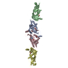
| ||||||||
|---|---|---|---|---|---|---|---|---|---|
| 1 |
| ||||||||
| Unit cell |
|
-Components
| #1: Protein | Mass: 29583.342 Da / Num. of mol.: 2 Source method: isolated from a genetically manipulated source Source: (gene. exp.) Zygosaccharomyces rouxii (strain ATCC 2623 / CBS 732 / NBRC 1130 / NCYC 568 / NRRL Y-229) (yeast)Strain: ATCC 2623 / CBS 732 / NBRC 1130 / NCYC 568 / NRRL Y-229 Gene: MMM1, ZYRO0B10274g / Production host:  #2: Protein | Mass: 23487.605 Da / Num. of mol.: 2 Source method: isolated from a genetically manipulated source Source: (gene. exp.) Strain: ATCC 204508 / S288c / Gene: MDM12, YOL009C / Production host: #3: Chemical | ChemComp-PO4 /   Mass: 94.971 Da / Num. of mol.: 6 / Source method: obtained synthetically / Formula: PO4 Mass: 94.971 Da / Num. of mol.: 6 / Source method: obtained synthetically / Formula: PO4 |
|---|
-Experimental details
-Experiment
| Experiment | Method: X-RAY DIFFRACTION / Number of used crystals: 1 |
|---|
- Sample preparation
| Crystal | Density Matthews: 4.73 Å3/Da / Density % sol: 74.01 % |
|---|---|
| Crystal grow | Temperature: 291 K / Method: vapor diffusion, hanging drop / Details: PEG 4000, HEPES, Ammonium sulfate |
-Data collection
| Diffraction | Mean temperature: 100 K |
|---|---|
| Diffraction source | Source: SYNCHROTRON / Site: PAL/PLS  / Beamline: 5C (4A) / Wavelength: 0.9795 Å / Beamline: 5C (4A) / Wavelength: 0.9795 Å |
| Detector | Type: DECTRIS PILATUS 6M / Detector: PIXEL / Date: Jul 21, 2017 |
| Radiation | Protocol: SINGLE WAVELENGTH / Monochromatic (M) / Laue (L): M / Scattering type: x-ray |
| Radiation wavelength | Wavelength: 0.9795 Å / Relative weight: 1 |
| Reflection | Resolution: 3.8→50 Å / Num. obs: 18459 / % possible obs: 99.8 % / Redundancy: 5.8 % / Rmerge(I) obs: 0.046 / Net I/σ(I): 27.5 |
| Reflection shell | Resolution: 3.8→3.87 Å |
- Processing
| Software |
| |||||||||||||||||||||||||||||||||||||||||||||||||||||||||||||||||||||||||||||||||||||||||||||||||||||||||||||||||||||||||||||
|---|---|---|---|---|---|---|---|---|---|---|---|---|---|---|---|---|---|---|---|---|---|---|---|---|---|---|---|---|---|---|---|---|---|---|---|---|---|---|---|---|---|---|---|---|---|---|---|---|---|---|---|---|---|---|---|---|---|---|---|---|---|---|---|---|---|---|---|---|---|---|---|---|---|---|---|---|---|---|---|---|---|---|---|---|---|---|---|---|---|---|---|---|---|---|---|---|---|---|---|---|---|---|---|---|---|---|---|---|---|---|---|---|---|---|---|---|---|---|---|---|---|---|---|---|---|---|
| Refinement | Method to determine structure: MOLECULAR REPLACEMENT Starting model: 5GYD Resolution: 3.799→39.142 Å / SU ML: 0.55 / Cross valid method: FREE R-VALUE / σ(F): 1.38 / Phase error: 33.23 / Stereochemistry target values: ML
| |||||||||||||||||||||||||||||||||||||||||||||||||||||||||||||||||||||||||||||||||||||||||||||||||||||||||||||||||||||||||||||
| Solvent computation | Shrinkage radii: 0.9 Å / VDW probe radii: 1.11 Å / Solvent model: FLAT BULK SOLVENT MODEL | |||||||||||||||||||||||||||||||||||||||||||||||||||||||||||||||||||||||||||||||||||||||||||||||||||||||||||||||||||||||||||||
| Refinement step | Cycle: LAST / Resolution: 3.799→39.142 Å
| |||||||||||||||||||||||||||||||||||||||||||||||||||||||||||||||||||||||||||||||||||||||||||||||||||||||||||||||||||||||||||||
| Refine LS restraints |
| |||||||||||||||||||||||||||||||||||||||||||||||||||||||||||||||||||||||||||||||||||||||||||||||||||||||||||||||||||||||||||||
| LS refinement shell |
| |||||||||||||||||||||||||||||||||||||||||||||||||||||||||||||||||||||||||||||||||||||||||||||||||||||||||||||||||||||||||||||
| Refinement TLS params. | Method: refined / Refine-ID: X-RAY DIFFRACTION
| |||||||||||||||||||||||||||||||||||||||||||||||||||||||||||||||||||||||||||||||||||||||||||||||||||||||||||||||||||||||||||||
| Refinement TLS group |
|
